
Computerized Axial Tomography Scan
A Computerized Axial Tomography scan, commonly known as a CT scan or CAT scan, is a sophisticated medical imaging technique that provides detailed cross-sectional images of the body. This non-invasive procedure plays a crucial role in diagnosing a wide range of medical conditions.
Key Takeaways
- A CT scan uses X-rays and computer processing to create detailed cross-sectional images of organs, soft tissues, bone, and blood vessels.
- It is a vital diagnostic tool for detecting conditions like tumors, internal injuries, fractures, and vascular diseases.
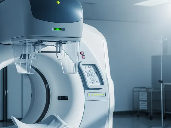
- The procedure involves lying on a table that slides into a large, donut-shaped scanner, often requiring a contrast agent for enhanced imaging.
- CT scans are generally safe, but involve exposure to ionizing radiation, which is carefully managed by medical professionals.
What is a Computerized Axial Tomography Scan?
A Computerized Axial Tomography scan is an advanced diagnostic imaging method that combines a series of X-ray images taken from different angles around your body. These images are then processed by a computer to create cross-sectional slices, or tomographic images, of bones, blood vessels, and soft tissues inside your body. Unlike a conventional X-ray, which provides a two-dimensional image, a CT scan offers a much more detailed, three-dimensional view of internal structures, allowing for precise identification of abnormalities.
How CT Scans Work and What They Reveal
Understanding how a CT scan works and what it shows involves appreciating its sophisticated technology. The process begins with a patient lying on a motorized table that moves through a large, donut-shaped machine called a gantry. Inside the gantry, an X-ray tube rotates around the patient, emitting narrow beams of X-rays. On the opposite side of the patient, detectors measure the X-ray beams that pass through the body.
The Imaging Process
As the X-ray tube and detectors rotate, they capture hundreds of thousands of individual X-ray measurements from various angles. A powerful computer then processes these measurements, assembling them into detailed cross-sectional images. Different tissues absorb X-rays at varying rates; for instance, bone appears white, while air appears black. This differential absorption allows the computer to construct clear images of organs, soft tissues, bone, and blood vessels. Sometimes, a contrast material—either swallowed, injected, or administered rectally—is used to highlight specific areas, such as blood vessels or the gastrointestinal tract, making them more visible on the images.
Conditions Diagnosed by CT
The detailed images produced by CT scans reveal a wide array of conditions that might not be visible on conventional X-rays. They are instrumental in detecting tumors, internal bleeding, blood clots, infections, and bone fractures. For example, CT scans can identify subtle fractures in complex bone structures like the spine or pelvis, which might be missed otherwise. According to the World Health Organization (WHO), medical imaging, including CT scans, is essential for diagnosing and monitoring numerous diseases, significantly contributing to global health outcomes by enabling early detection and treatment.
Medical Uses and the CAT Scan Procedure
The purpose and uses of CAT scans in medical diagnosis are extensive, making them indispensable in modern healthcare. From emergency situations to routine check-ups, CT scans provide critical information that guides treatment decisions. They are frequently used to assess injuries from trauma, diagnose cancers, evaluate cardiovascular conditions, and guide biopsies or other minimally invasive procedures.
Common Diagnostic Applications
CT scans are particularly valuable in several medical fields. In oncology, they help stage cancer, monitor treatment effectiveness, and detect recurrence. For cardiovascular health, they can identify blockages in arteries (CT angiography) or assess the extent of heart disease. In emergency medicine, CT scans quickly identify internal injuries, such as those to the brain, lungs, or abdominal organs, following accidents. They are also used to detect kidney stones, appendicitis, and various musculoskeletal disorders. The ability to visualize intricate internal structures makes CT an unparalleled tool for comprehensive diagnostic assessment.
Preparing for Your CT Scan
Understanding the Computerized Axial Tomography procedure involves knowing what to expect during preparation and the scan itself. Patients are typically advised to wear comfortable, loose-fitting clothing and may be asked to remove metal objects like jewelry, eyeglasses, or dentures, as these can interfere with the X-ray images. Depending on the area being scanned and whether a contrast agent is required, patients might be instructed to fast for a few hours before the procedure. If a contrast agent is used, you may feel a warm flush or a metallic taste in your mouth, which is normal and temporary. The scan itself is painless and usually takes between 10 to 30 minutes, during which you will need to lie still to ensure clear images.


