
Compound Nevus
A compound nevus is a common type of mole characterized by the presence of nevus cells in both the epidermis and the dermis. While generally benign, understanding their characteristics and potential need for evaluation is crucial for skin health.
Key Takeaways
- A Compound Nevus is a common mole with melanocytes in both epidermal and dermal layers of the skin.
- They typically appear as raised, dome-shaped lesions, often flesh-colored to dark brown, and are usually benign.
- Causes include genetic factors and sun exposure, with professional evaluation being vital for differential diagnosis against melanoma.
- Removal options, such as surgical excision, are considered for cosmetic reasons, irritation, or suspicion of malignancy.
- The prognosis for a compound nevus is excellent, as they are almost always non-cancerous.
What is a Compound Nevus?
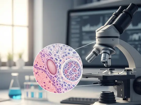
A Compound Nevus is a type of melanocytic nevus, commonly known as a mole, where the nevus cells (melanocytes) are found in both the epidermal-dermal junction and within the dermis itself. This dual location gives them their characteristic raised appearance, distinguishing them from other types of nevi. These moles are very common, with most adults having several nevi on their skin. According to the American Academy of Dermatology, it is normal for adults to have between 10 and 40 moles, many of which are compound nevi.
Understanding the distinction between different types of nevi is important for diagnosis. Here’s a comparison of a compound nevus with other common types:
| Nevus Type | Location of Melanocytes | Typical Appearance |
|---|---|---|
| Junctional Nevus | Epidermal-dermal junction | Flat, well-defined, dark brown to black |
| Compound Nevus | Epidermal-dermal junction AND dermis | Raised, dome-shaped, often light brown to dark brown, sometimes flesh-colored |
| Intradermal Nevus | Dermis only | Raised, dome-shaped, often flesh-colored or light brown, may have hairs |
The term “compound nevus vs intradermal nevus” highlights a key difference in melanocyte distribution, influencing their clinical presentation. While both can be raised, the presence of junctional components in a compound nevus can sometimes lead to a darker, more variegated appearance compared to a purely intradermal nevus.
Compound Nevus Symptoms, Causes, and Differential Diagnosis
The typical compound nevus symptoms causes are generally benign and relate to their appearance. They often present as slightly raised, dome-shaped lesions, ranging in color from flesh-toned to light or dark brown. They can be smooth or warty in texture and may sometimes contain hairs. The size usually remains stable over time, typically less than 6 millimeters in diameter. While most compound nevi are harmless, any changes in size, shape, color, or sensation (itching, bleeding) warrant medical evaluation.
The exact causes of compound nevi are multifactorial. Genetic predisposition plays a significant role, meaning individuals with a family history of moles may be more prone to developing them. Additionally, exposure to ultraviolet (UV) radiation from the sun or tanning beds is a known contributing factor to the development of various types of nevi. Hormonal changes, such as those occurring during puberty or pregnancy, can also influence the appearance and number of moles.
Differential diagnosis is critical to distinguish a benign compound nevus from more serious conditions, particularly melanoma. Dermatologists use the “ABCDE” rule to assess moles for suspicious characteristics:
- Asymmetry: One half of the mole does not match the other.
- Border: The edges are irregular, ragged, notched, or blurred.
- Color: The color is not uniform and may include shades of brown, black, tan, white, red, or blue.
- Diameter: The mole is larger than 6 millimeters (about the size of a pencil eraser).
- Evolving: The mole is changing in size, shape, color, or elevation, or new symptoms like bleeding, itching, or crusting appear.
Any mole exhibiting these features should be promptly examined by a dermatologist for accurate diagnosis and appropriate management.
Compound Nevus Removal Options and Prognosis
When considering compound nevus removal options, the decision is typically based on cosmetic concerns, irritation from clothing or shaving, or, most importantly, suspicion of malignancy. If a mole exhibits any atypical features or changes, removal for histopathological examination is often recommended to rule out melanoma.
Common removal techniques include:
- Surgical Excision: This involves cutting out the entire mole and a small margin of surrounding healthy skin, followed by stitching the wound closed. This method ensures complete removal and allows for thorough microscopic examination of the tissue.
- Shave Excision: For raised moles, a shave excision can be performed where the mole is shaved off flush with the skin surface using a scalpel. This is often used for cosmetic reasons or when malignancy is less suspected, but it may not remove the entire nevus if it extends deep into the dermis.
- Punch Biopsy: A circular tool is used to remove a small, cylindrical piece of the mole. This is often done for diagnostic purposes, with the option for complete excision if needed.
Other methods, such as laser removal, are generally not recommended for pigmented lesions due to the inability to obtain tissue for pathological analysis, which is crucial for ruling out melanoma. The prognosis for a compound nevus is excellent, as they are benign lesions and do not typically pose a health risk. Once removed, recurrence is rare if the excision was complete. However, individuals with numerous moles or a history of atypical nevi should continue regular self-skin exams and professional dermatological check-ups to monitor for new or changing lesions.


