Capillary Malformation
Capillary Malformation is a type of vascular birthmark characterized by abnormally dilated capillaries in the skin. These birthmarks are typically present at birth and can vary in size and appearance.
Key Takeaways
- Capillary Malformation is a common vascular birthmark resulting from dilated capillaries.
- It presents as a flat, pink to reddish-purple patch on the skin, often growing proportionally with the individual.
- Diagnosis is primarily clinical, based on the characteristic appearance of the lesion.
- The underlying causes of capillary malformation are typically sporadic genetic mutations.
- Treatment often involves laser therapy, particularly pulsed dye laser (PDL), to lighten the lesion.
What is Capillary Malformation?
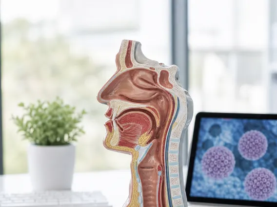
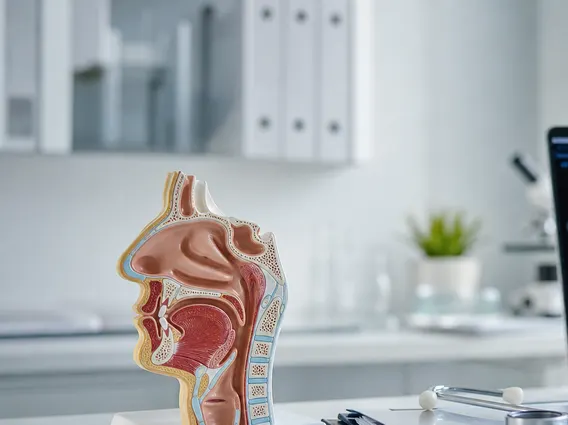
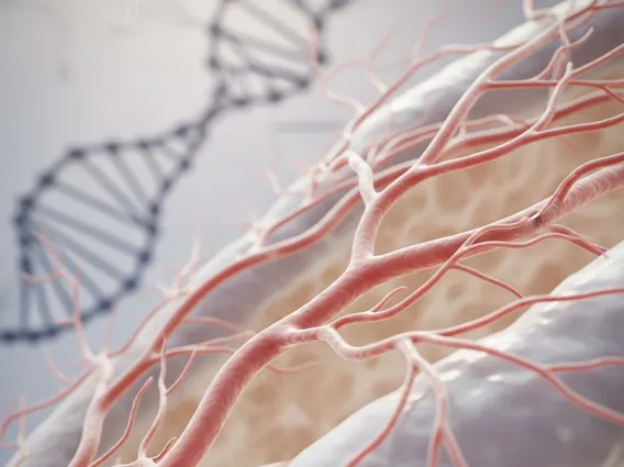
A Capillary Malformation is a common type of vascular birthmark, often referred to as a port-wine stain. It is characterized by an area of skin where the small blood vessels (capillaries) are abnormally dilated and numerous, leading to a distinctive discoloration. These malformations are present from birth and do not fade over time; in fact, they may darken and thicken with age. They can appear anywhere on the body, though they are most common on the face and neck. According to the American Academy of Dermatology, capillary malformations affect approximately 0.3% of newborns, making them one of the more prevalent types of vascular birthmarks.
Unlike some other birthmarks, capillary malformations are not typically raised or bumpy at birth. They are flat patches that can range in color from light pink to deep reddish-purple, depending on the depth and density of the affected capillaries. While most capillary malformations are isolated skin findings, some can be associated with underlying syndromes, such as Sturge-Weber syndrome when located on the face, particularly involving the forehead and eyelid, or Klippel-Trenaunay syndrome when affecting a limb.
Identifying Capillary Malformation: Symptoms and Diagnosis
The primary capillary malformation symptoms are visual: a flat, discolored patch of skin that is present at birth. These lesions typically grow proportionally with the child and do not resolve spontaneously. Over time, particularly in adulthood, the affected skin may become thicker, develop small bumps (nodules), and deepen in color. The texture can also change, becoming more uneven or pebbly.
Diagnosis of a capillary malformation is usually clinical, based on a thorough physical examination and the characteristic appearance of the birthmark. A dermatologist or pediatrician can typically identify the condition without the need for invasive tests. In cases where the malformation is extensive, involves certain areas of the face (like the eyelid or forehead), or is associated with other symptoms, further evaluation may be necessary to rule out associated syndromes. This might include:
- Neurological imaging (e.g., MRI) to check for brain involvement in suspected Sturge-Weber syndrome.
- Ophthalmological examination to assess for glaucoma, which can be associated with facial capillary malformations.
- Limb measurements or imaging to check for limb overgrowth in suspected Klippel-Trenaunay syndrome.
Early and accurate diagnosis is important for appropriate monitoring and to discuss potential treatment options.
Causes and Treatment Options for Capillary Malformation
The exact causes of capillary malformation are not fully understood, but they are generally believed to result from sporadic somatic mutations. This means the genetic change occurs after conception in a single cell and is not inherited from parents. Research has identified mutations in genes such as GNAQ and GNA11 as being linked to the development of these malformations. These mutations lead to abnormal signaling pathways that affect the development and function of capillaries, causing them to dilate and become more numerous in the affected skin area. Most capillary malformations occur randomly and are not passed down through families.
Regarding capillary malformation treatment info, the primary and most effective treatment is pulsed dye laser (PDL) therapy. This laser specifically targets the hemoglobin within the red blood cells in the dilated capillaries, causing them to heat up and coagulate, without significantly damaging the surrounding skin. The body then reabsorbs these damaged vessels, leading to a lightening of the birthmark. Multiple treatment sessions are typically required, often starting in infancy, to achieve optimal results. Early treatment can sometimes lead to better outcomes and may prevent the thickening and darkening that can occur later in life.
Other management strategies may include:
- Cosmetic camouflage makeup to cover the lesion.
- Surgical excision for very small, localized lesions or for thickened, nodular areas that do not respond well to laser therapy.
- Ongoing monitoring for any associated complications, such as glaucoma or limb overgrowth, especially in syndromic cases.
It is important to consult with a dermatologist or a specialist in vascular anomalies to determine the most appropriate treatment plan, as individual responses to therapy can vary.