
Key Takeaways
- Retinoblastoma is a rare childhood eye cancer, often presenting before the age of five.
- The most common initial sign is leukocoria, a white reflex in the pupil, often noticed in photographs.
- Genetic mutations, particularly in the RB1 gene, are the primary causes and risk factors of retinoblastoma.
- Early detection of retinoblastoma signs through regular pediatric eye exams is crucial for successful treatment and favorable outcomes.
- Modern retinoblastoma treatment options for children offer high survival rates and improved chances of vision preservation.
Understanding Retinoblastoma Eye Cancer
Definition and types
Retinoblastoma eye cancer is a malignant tumor that develops from immature cells of the retina. It is the most common primary intraocular malignancy in childhood, typically diagnosed in children under five years old. This cancer can affect one eye (unilateral retinoblastoma) or both eyes (bilateral retinoblastoma).
Globally, retinoblastoma affects approximately 1 in 15,000 to 1 in 20,000 live births, according to the World Health Organization. While rare, its potential for rapid growth and spread makes understanding its nature critical. The disease is categorized into two main forms: heritable (germline mutation) and non-heritable (somatic mutation), which influences the risk of recurrence and secondary cancers.
How it develops
The development of retinoblastoma is primarily linked to mutations in the RB1 gene, a tumor suppressor gene. When this gene is mutated or missing, retinal cells can grow uncontrollably, leading to tumor formation. These tumors originate in the neurosensory retina, specifically from retinoblasts, which are immature retinal cells.
As the tumor grows, it can fill the eye, detach the retina, and in advanced stages, spread beyond the eye to other parts of the body, including the brain and bone marrow. The speed of development varies, but early detection is key to preventing metastasis and ensuring effective intervention.
Recognizing Early Signs in Infants
Common visual indicators
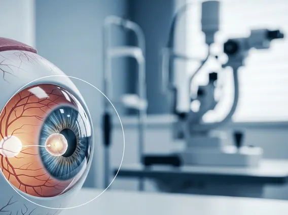
Recognizing retinoblastoma symptoms in infants is paramount for timely diagnosis and treatment. The most common and often first noticeable sign is leukocoria, also known as the “cat’s eye reflex.” This appears as a white glow in the pupil when light, such as a camera flash, hits the eye, instead of the normal red reflex.
Other significant visual indicators include strabismus (crossed eyes or “lazy eye”), which occurs if the tumor affects central vision, causing the eye to wander. Parents might also notice a red or irritated eye that persists without infection, or a difference in the size of the pupils (anisocoria). In some cases, children may exhibit poor vision or nystagmus (involuntary eye movements).
When to seek medical attention
Early detection of retinoblastoma signs significantly improves the prognosis. Parents and caregivers should be vigilant for any unusual changes in a child’s eyes or vision. If leukocoria is observed, even in a single photograph, immediate medical attention from a pediatrician or ophthalmologist is crucial. Similarly, persistent strabismus, unexplained eye redness, or any concerns about a child’s visual development warrant prompt evaluation.
Routine well-child check-ups often include a red reflex test, which can sometimes identify retinoblastoma before other symptoms become apparent. However, parents should not wait for these appointments if they notice any suspicious signs, as rapid assessment can be life-saving and vision-preserving.
Causes, Risk Factors, and Diagnosis
Genetic and hereditary factors
The primary causes and risk factors of retinoblastoma are genetic. Approximately 60% of cases are non-heritable, meaning the RB1 gene mutation occurs spontaneously in a single retinal cell. The remaining 40% are heritable, caused by a germline mutation in the RB1 gene, which is present in all cells of the body.
Children with heritable retinoblastoma often develop tumors in both eyes (bilateral) and are at a higher risk of developing other cancers later in life, such as osteosarcoma or pinealoblastoma. Genetic counseling and testing are vital for families with a history of retinoblastoma to assess risk and guide screening protocols for at-risk infants.
Diagnostic procedures
The process for retinoblastoma diagnosis is thorough and typically begins with a comprehensive dilated eye examination performed by an ophthalmologist, often under general anesthesia for infants and young children. This allows for a detailed view of the retina and the identification of any tumors.
Further diagnostic procedures include:
- Ocular Ultrasound: Uses sound waves to create images of the eye, helping to confirm the presence of a tumor and detect calcifications, which are common in retinoblastoma.
- Magnetic Resonance Imaging (MRI): Provides detailed images of the eye and surrounding structures, crucial for assessing tumor size, extent, and whether it has spread outside the eye or to the brain.
- Genetic Testing: Performed on blood or tumor tissue to identify RB1 gene mutations, which helps determine if the retinoblastoma is heritable and informs family screening.
- Lumbar Puncture and Bone Marrow Biopsy: May be conducted in advanced cases to check for metastasis to the central nervous system or bone marrow, though this is rare at initial diagnosis.
These diagnostic tools collectively provide a clear picture of the tumor’s characteristics, enabling the medical team to stage the disease and formulate an appropriate treatment plan.
Retinoblastoma Treatment & Patient Prognosis
Modern therapeutic approaches
Various retinoblastoma treatment options for children are available, tailored to the tumor’s size, location, and whether it affects one or both eyes. The primary goals are to eradicate the cancer, preserve the child’s life, save the eye, and maximize vision.
Treatment modalities often include a combination of the following:
- Systemic Chemotherapy: Used to shrink large tumors before local treatment or to treat metastatic disease.
- Intra-arterial Chemotherapy: Delivers chemotherapy directly to the eye via a catheter, minimizing systemic side effects.
- Intravitreal Chemotherapy: Injects chemotherapy directly into the vitreous gel of the eye for tumors that have spread within the eye.
- Focal Therapies:
- Laser Photocoagulation: Uses a laser to destroy small tumors by heating them.
- Cryotherapy: Freezes and thaws small tumors to destroy cancer cells.
- Thermotherapy: Uses heat to destroy cancer cells, often combined with chemotherapy.
- Radiation Therapy:
- Brachytherapy (Plaque Radiotherapy): A small radioactive disc is temporarily sewn onto the eye over the tumor.
- External Beam Radiation: Uses high-energy rays to destroy cancer cells, typically reserved for advanced cases or when other treatments fail, due to potential long-term side effects.
- Enucleation: Surgical removal of the eye, usually performed for very large tumors, advanced disease, or when vision cannot be saved, to prevent the spread of cancer.
The choice of treatment is highly individualized, with a multidisciplinary team of specialists collaborating to achieve the best possible outcome for each child.
Long-term outlook for patients
The prognosis for retinoblastoma patients has dramatically improved over the past few decades, particularly in developed countries. With early diagnosis and appropriate treatment, the survival rate for retinoblastoma is over 95%, according to the American Cancer Society. However, the long-term outlook also involves considerations beyond survival, such as vision preservation and the risk of secondary cancers.
Children who have undergone treatment require lifelong follow-up care, including regular eye examinations to monitor for recurrence and to manage any visual impairments. For those with heritable retinoblastoma, continued surveillance for secondary cancers is crucial due to their increased genetic predisposition. Advances in treatment continue to enhance both survival rates and the quality of life for children affected by this rare eye cancer.
Retinoblastoma FAQs
The most common initial sign of retinoblastoma is leukocoria, often referred to as the “cat’s eye reflex.” This appears as an abnormal white glow in the pupil, especially noticeable in photographs taken with a flash, instead of the typical red reflex. Other early indicators can include strabismus (crossed eyes), persistent eye redness, or a noticeable difference in pupil size. Any of these signs warrant immediate medical evaluation by a pediatrician or ophthalmologist to ensure timely diagnosis and intervention.
No, retinoblastoma is not always hereditary. Approximately 40% of cases are hereditary, meaning they result from a germline mutation in the RB1 gene that is present in all cells and can be passed down. These children often develop tumors in both eyes. The remaining 60% of cases are non-heritable, caused by a spontaneous mutation in a single retinal cell. Genetic testing can determine if the retinoblastoma is hereditary, which is crucial for family counseling and screening of at-risk siblings.
The survival rate for retinoblastoma patients, especially with early detection and access to modern medical care, is very high. In developed countries, the survival rate exceeds 95%. This impressive statistic is a testament to advancements in diagnostic techniques and a range of effective treatment options, including chemotherapy, focal therapies, and surgery. While survival is excellent, the focus also remains on preserving vision and managing potential long-term effects, including the risk of secondary cancers in hereditary cases.