
Bronchial Washing
Bronchial washing is a diagnostic medical procedure used to collect fluid and cells from the airways of the lungs. This technique helps healthcare professionals identify various respiratory conditions by examining the collected samples.
Key Takeaways
- Bronchial washing is a diagnostic procedure involving the collection of fluid from lung airways to detect respiratory diseases.
- It is primarily used to diagnose infections, inflammation, and certain cancers within the lungs.
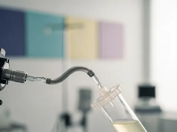
- The procedure involves a bronchoscope, where saline is instilled and then suctioned out to gather samples.
- Potential risks are generally low but can include bleeding, infection, or temporary breathing difficulties.
- Interpreting the results helps guide treatment plans for conditions like pneumonia, tuberculosis, and lung cancer.
What is Bronchial Washing?
Bronchial washing is a medical technique performed during a bronchoscopy, where a sterile saline solution is introduced into the bronchial tubes and then immediately suctioned back out. This process collects cells, microorganisms, and other substances present in the airways, providing valuable samples for laboratory analysis. It is a crucial component of the broader bronchial washing procedure, enabling direct assessment of the lower respiratory tract.
Purpose of Bronchial Washing
The primary bronchial washing purpose is to obtain diagnostic samples from the lungs when other, less invasive tests have been inconclusive or are not suitable. It allows for the identification of pathogens, abnormal cells, and inflammatory markers directly from the site of concern. This direct sampling is vital for accurate diagnosis and effective treatment planning, minimizing the need for more invasive surgical biopsies in many cases.
Conditions Diagnosed by Bronchial Washing
Bronchial washing is instrumental in diagnosing a wide range of pulmonary conditions. According to the World Health Organization (WHO), respiratory diseases are a leading cause of morbidity and mortality globally, making accurate diagnostic tools like bronchial washing essential. Conditions commonly diagnosed include:
- Infections: Bacterial, viral, fungal, or parasitic infections, including pneumonia, tuberculosis, and atypical mycobacterial infections.
- Inflammatory Conditions: Such as sarcoidosis or certain types of pneumonitis, where inflammatory cells or specific markers can be identified.
- Cancers: Detection of malignant cells, particularly in cases of suspected lung cancer, helping to determine the type and stage of the cancer.
- Bleeding Disorders: Identifying the source and nature of bleeding within the airways.
The Bronchial Washing Procedure
Understanding how is bronchial washing performed involves several key stages, from initial preparation to the actual collection of samples. The procedure is typically carried out in a hospital or specialized clinic setting by a pulmonologist or an interventional bronchoscopist.
Preparation for the Procedure
Before the procedure, patients are usually advised to fast for several hours. They may also undergo preliminary tests such as blood work, chest X-rays, or CT scans. Patients will receive a local anesthetic to numb the throat and often a sedative to help them relax. This ensures comfort and minimizes discomfort during the procedure.
Steps of Bronchial Washing
The actual steps of the bronchial washing procedure are meticulous and carefully executed:
- Bronchoscope Insertion: A thin, flexible tube called a bronchoscope, equipped with a light and camera, is gently inserted through the nose or mouth, down the trachea, and into the bronchial tubes.
- Airway Visualization: The physician navigates the bronchoscope to the specific area of the lung suspected of disease, visualizing the airways on a monitor.
- Saline Instillation: A small amount of sterile saline solution (typically 20-60 ml) is instilled into the targeted bronchial segment through the bronchoscope.
- Sample Collection: The saline, now mixed with cells and other substances from the airway lining, is immediately suctioned back into a sterile collection trap. This process may be repeated multiple times to ensure an adequate sample.
- Bronchoscope Withdrawal: Once sufficient samples are collected, the bronchoscope is carefully withdrawn.
Potential Risks and Side Effects
While generally safe, the bronchial washing purpose and risks must be considered. The risks associated with the procedure are typically low but can include:
- Bleeding: Minor bleeding can occur, especially if a biopsy is also performed.
- Infection: Though rare, there’s a small risk of introducing infection into the lungs.
- Pneumothorax: A collapsed lung, which is a very rare complication.
- Bronchospasm: Temporary tightening of the airways, leading to shortness of breath.
- Sore Throat/Hoarseness: Common temporary side effects due to the bronchoscope.
Patients are monitored closely after the procedure to ensure a smooth recovery and address any immediate concerns.
Interpreting Bronchial Washing Results
The bronchial washing results interpretation is a critical step performed by pathologists and physicians. The collected fluid samples are sent to a laboratory for various analyses, including cytology (examination of cells), microbiology (culture for bacteria, fungi, viruses), and sometimes molecular tests.
What Abnormal Results May Indicate
Abnormal findings from bronchial washing can point to several underlying health issues. The specific indicators help guide further diagnostic steps and treatment strategies:
| Abnormal Finding | Potential Indication |
|---|---|
| Presence of specific bacteria, fungi, or viruses | Bacterial pneumonia, fungal infection (e.g., aspergillosis), viral bronchitis, or tuberculosis. |
| Malignant cells | Lung cancer (e.g., adenocarcinoma, squamous cell carcinoma). |
| Increased inflammatory cells (e.g., neutrophils, eosinophils) | Inflammatory lung diseases, allergic reactions, or certain types of pneumonia. |
| Hemosiderin-laden macrophages | Recent or chronic bleeding in the lungs. |
| Specific protein markers | Certain interstitial lung diseases or autoimmune conditions. |
A comprehensive interpretation considers these findings in conjunction with the patient’s clinical history, symptoms, and other imaging results to arrive at a definitive diagnosis and formulate an appropriate treatment plan.


