Biopsy
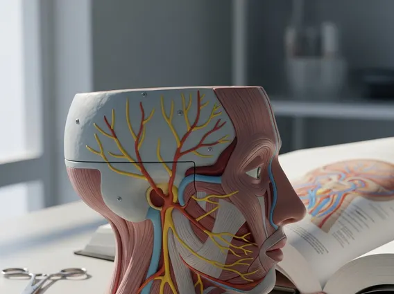
A biopsy is a medical procedure involving the removal of tissue or cells for examination under a microscope. It is a critical diagnostic tool used to determine the presence or extent of a disease, providing essential information for treatment planning.

Key Takeaways
- A biopsy is the removal of tissue for microscopic examination, crucial for diagnosing various conditions.
- It is primarily performed to confirm or rule out diseases like cancer, infections, or inflammatory conditions.
- Different types of biopsy procedures exist, chosen based on the suspected condition and location, such as needle, excisional, or endoscopic biopsies.
- Results are interpreted by a pathologist, providing a definitive diagnosis that guides further medical management.
- Understanding your biopsy results is vital for informed discussions with your healthcare provider about treatment options.
What is Biopsy and Why It’s Performed
A biopsy is a diagnostic medical procedure where a small sample of tissue or cells is extracted from the body for detailed examination. This examination, typically performed by a pathologist, helps to identify abnormalities that cannot be fully understood through imaging tests alone. The primary purpose of what is a biopsy procedure is to obtain definitive information about a suspicious area.
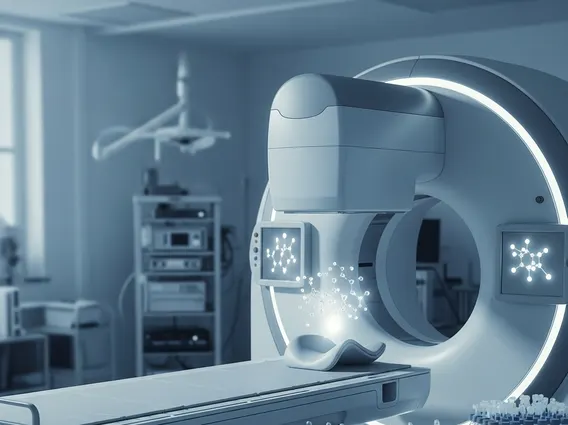
The question of why is a biopsy performed often arises when other diagnostic methods, such as X-rays, CT scans, MRIs, or blood tests, indicate a potential issue that requires further investigation. For instance, if a lump is detected during a physical exam or an abnormal growth appears on an imaging scan, a biopsy can determine if it is benign (non-cancerous) or malignant (cancerous). According to the American Cancer Society, biopsies are the only definitive way to diagnose most cancers, playing a pivotal role in early detection and treatment planning.
Types of Biopsy Procedures
There are several types of biopsy and their uses, each selected based on the location of the suspicious tissue, its accessibility, and the suspected condition. The choice of procedure aims to obtain an adequate sample with minimal invasiveness and discomfort for the patient.
Common biopsy types include:
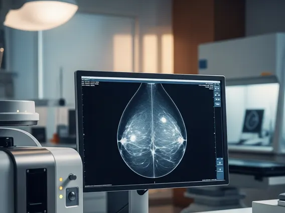
- Needle Biopsy: This involves using a special needle to extract tissue. It can be a fine-needle aspiration (FNA) for fluid or small cell samples, or a core needle biopsy (CNB) for larger tissue samples. It’s often guided by imaging techniques like ultrasound or CT scans, particularly for deep-seated lesions in organs like the liver, lung, or breast.
- Excisional Biopsy: In this procedure, the entire suspicious lump or abnormal area is removed, along with a margin of surrounding healthy tissue. It is frequently used for skin lesions or small tumors that are easily accessible.
- Incisional Biopsy: Similar to an excisional biopsy, but only a portion of a larger tumor or lesion is removed for diagnosis. This is done when removing the entire mass is not feasible or necessary for initial diagnosis.
- Endoscopic Biopsy: Performed during an endoscopy, colonoscopy, or bronchoscopy, where a flexible tube with a camera is inserted into the body (e.g., gastrointestinal tract, lungs). Small forceps at the end of the scope are used to snip off tiny tissue samples from the lining of these organs.
- Bone Marrow Biopsy: Involves taking a sample of bone marrow (the spongy tissue inside bones) to diagnose blood disorders, certain cancers, or infections. It is typically taken from the hip bone.
Interpreting Your Biopsy Results
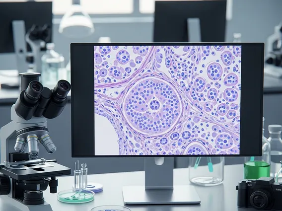
Once a tissue sample is collected, it is sent to a pathology laboratory for analysis. A pathologist, a doctor specializing in diagnosing diseases by examining tissues and body fluids, processes the sample and views it under a microscope. They look for abnormal cells, tissue architecture changes, or specific markers that indicate disease. The process of biopsy results interpretation can take several days to a week, depending on the complexity of the case and the tests required.
The pathologist then generates a detailed report, which is sent to the referring physician. This report will typically include a macroscopic description (what the tissue looked like to the naked eye), a microscopic description (what was seen under the microscope), and a final diagnosis. Your doctor will then discuss these results with you, explaining what they mean for your health and outlining any necessary next steps, such as further tests or treatment plans. It is crucial to ask questions and ensure you understand the diagnosis and its implications for your care.