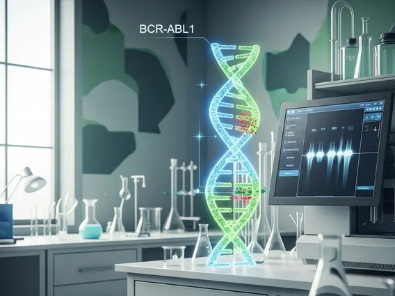
BCRABL1 Fusion Gene
The BCRABL1 fusion gene is a critical genetic abnormality associated with several types of leukemia. Its presence significantly influences disease progression, diagnosis, and treatment strategies in affected individuals.
Key Takeaways
- The BCR-ABL1 fusion gene results from a specific chromosomal translocation, often known as the Philadelphia chromosome.
- It produces an abnormal protein with unregulated tyrosine kinase activity, leading to uncontrolled cell growth.
- This gene is primarily associated with Chronic Myeloid Leukemia (CML) and also found in some cases of Acute Lymphoblastic Leukemia (ALL) and Acute Myeloic Leukemia (AML).
- Detection of BCR-ABL1 is crucial for diagnosis, prognosis, and guiding targeted therapies, such as tyrosine kinase inhibitors (TKIs).
- Various methods, including cytogenetics, FISH, and RT-qPCR, are used to identify and monitor the BCR-ABL1 fusion gene.
What is the BCRABL1 Fusion Gene (BCR-ABL1)?
The BCR-ABL1 fusion gene, also commonly referred to as the Philadelphia chromosome, is a genetic abnormality that arises from a reciprocal translocation between chromosome 9 and chromosome 22. This specific rearrangement results in the fusion of the breakpoint cluster region (BCR) gene on chromosome 22 with the Abelson murine leukemia viral oncogene homolog 1 (ABL1) gene on chromosome 9. The resulting fusion gene, BCR-ABL1, produces an abnormal protein with continuously active tyrosine kinase activity.
This constitutively active tyrosine kinase drives uncontrolled cell proliferation and inhibits programmed cell death (apoptosis), leading to the accumulation of abnormal white blood cells. Understanding the mechanism by which the BCR-ABL1 fusion gene causes and effects cellular changes is fundamental to comprehending the pathogenesis of associated leukemias. The presence of this fusion gene is a hallmark diagnostic marker and a primary target for specific therapeutic interventions.
Impact and Associated Conditions
The primary condition associated with the BCR-ABL1 fusion gene is Chronic Myeloid Leukemia (CML), where it is found in over 95% of cases. Its presence is a defining characteristic of CML, driving the disease from its chronic phase through potential accelerated and blast phases. Beyond CML, the BCR-ABL1 fusion gene can also be detected in a subset of patients with Acute Lymphoblastic Leukemia (ALL), particularly in adults, and less frequently in Acute Myeloid Leukemia (AML). In these acute leukemias, the presence of BCR-ABL1 often indicates a more aggressive disease course and can influence treatment choices.
The continuous signaling from the abnormal BCR-ABL1 protein leads to a cascade of cellular events that promote leukemic cell survival and growth. This impact makes the BCR-ABL1 fusion gene a highly significant prognostic indicator. For instance, in CML, patients with the BCR-ABL1 fusion gene typically respond well to targeted therapies known as tyrosine kinase inhibitors (TKIs), which specifically block the activity of the BCR-ABL1 protein. The success of these targeted treatments has transformed CML from a fatal disease into a manageable chronic condition for many patients. According to the World Health Organization (WHO), the identification of this fusion gene is critical for accurate diagnosis and for guiding personalized treatment strategies in these hematological malignancies.
Detection Methods for BCRABL1
Accurate detection of the BCRABL1 fusion gene detection methods are essential for the diagnosis, prognosis, and monitoring of patients with suspected or confirmed CML and other associated leukemias. Several sophisticated molecular and cytogenetic techniques are employed for this purpose, each offering different levels of sensitivity and specificity. These methods allow clinicians to identify the presence of the gene, quantify its expression, and track treatment response over time.
Common BCRABL1 fusion gene detection methods include:
- Cytogenetics (Karyotyping): This traditional method involves analyzing chromosomes under a microscope to identify the Philadelphia chromosome (Ph), which is the visible manifestation of the t(9;22) translocation. While effective for initial detection, it may not be sensitive enough to detect small populations of cells with the fusion gene or cryptic translocations.
- Fluorescence In Situ Hybridization (FISH): FISH uses fluorescently labeled DNA probes that bind to specific sequences on chromosomes. It can detect the BCR-ABL1 fusion gene even when the Philadelphia chromosome is not clearly visible by standard cytogenetics, offering higher sensitivity.
- Quantitative Reverse Transcription Polymerase Chain Reaction (RT-qPCR): This is the most sensitive method for detecting and quantifying BCR-ABL1 messenger RNA (mRNA) levels. RT-qPCR is crucial for monitoring minimal residual disease (MRD) during treatment, allowing clinicians to assess the effectiveness of therapies and predict potential relapse. It can detect very low levels of the fusion transcript, making it invaluable for long-term patient management.
The choice of detection method often depends on the clinical context, including initial diagnosis, confirmation, or ongoing monitoring of treatment efficacy. Regular monitoring of BCR-ABL1 levels, particularly with RT-qPCR, helps guide therapeutic adjustments and ensures optimal patient outcomes.