
Clark Levels
Clark Levels represent a historical classification system used in dermatology and oncology to assess the depth of invasion of melanoma into the skin layers. This system provides crucial insights into the tumor’s aggressiveness and helps guide treatment strategies, though it has largely been supplemented by other metrics in modern staging.
Key Takeaways
- Clark Levels classify melanoma based on its depth of penetration into the skin.
- There are five distinct levels (I-V), ranging from melanoma confined to the epidermis to invasion into subcutaneous fat.
- Higher Clark Levels indicate deeper invasion and are generally associated with a less favorable prognosis.
- While historically significant for melanoma staging, Clark Levels have largely been superseded by Breslow depth due to its greater prognostic accuracy.
- Understanding Clark Levels still provides valuable context for assessing tumor biology and guiding clinical decisions.
What are Clark Levels: Understanding Melanoma Depth
Clark Levels refer to a system developed by Dr. Wallace H. Clark Jr. in 1969 to categorize the depth of melanoma invasion into the skin. This classification was groundbreaking for its time, providing a standardized method to assess how deeply a melanoma had penetrated the various layers of the skin. The primary purpose of this system is to evaluate the extent of tumor growth vertically, which is a critical factor in determining prognosis and treatment.
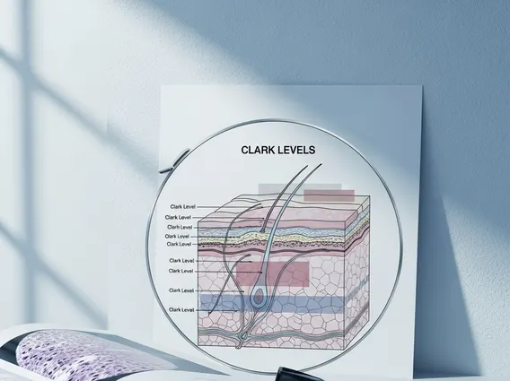
The skin is composed of several layers, and the Clark Levels delineate invasion relative to these anatomical structures. These layers include the epidermis (the outermost layer), the papillary dermis (the superficial part of the dermis), the reticular dermis (the deeper, thicker part of the dermis), and the subcutaneous fat (the layer beneath the dermis). By identifying which of these layers the melanoma has reached, clinicians can gain insight into the potential for the cancer to spread.
Clark Level Classification Explained for Staging
The Clark level classification explained system divides melanoma invasion into five distinct levels, each corresponding to a specific anatomical depth within the skin. While modern staging predominantly relies on Breslow depth (a quantitative measurement in millimeters), Clark Levels still offer a qualitative understanding of the tumor’s vertical growth. This system was historically vital for clark levels melanoma staging, helping clinicians predict the likelihood of metastasis and recurrence.
Here is a detailed breakdown of each Clark Level:
| Clark Level | Description of Invasion |
|---|---|
| Level I | Melanoma confined to the epidermis (melanoma in situ). It has not invaded the dermis. |
| Level II | Melanoma invades the papillary dermis (the superficial layer of the dermis). |
| Level III | Melanoma fills and expands the papillary dermis, reaching the interface with the reticular dermis, but does not invade the reticular dermis itself. |
| Level IV | Melanoma invades the reticular dermis (the deeper, thicker layer of the dermis). |
| Level V | Melanoma invades the subcutaneous fat (the fatty tissue beneath the dermis). |
Understanding these levels helps illustrate the progression of melanoma from a superficial lesion to a more deeply invasive tumor. The deeper the invasion, the higher the Clark Level, generally indicating a more advanced stage of the disease.
Clinical Significance and Prognostic Value
The clinical significance of Clark Levels lies in their correlation with prognosis. Generally, a higher Clark Level indicates a greater risk of regional lymph node involvement and distant metastasis, leading to a less favorable prognosis. For instance, a melanoma at Clark Level I (in situ) has an excellent prognosis with appropriate treatment, whereas a melanoma at Clark Level V carries a significantly higher risk of recurrence and spread.
While Clark Levels provided foundational insights into clark levels skin cancer depth, their use as the primary prognostic indicator has been largely replaced by Breslow depth. Breslow depth, which measures the tumor’s vertical thickness in millimeters from the granular layer of the epidermis to the deepest point of invasion, has proven to be a more accurate and reproducible predictor of patient outcomes. However, Clark Levels still offer valuable qualitative information, particularly in cases where Breslow depth might be difficult to measure precisely or for historical comparison.
Accurate assessment of tumor depth is paramount for effective treatment planning. According to the American Cancer Society, early detection and precise staging, including the evaluation of tumor depth, are crucial factors that significantly improve survival rates for melanoma patients. Therefore, while Breslow depth is the current gold standard, the conceptual understanding provided by Clark Levels remains an important part of dermatological and oncological education regarding melanoma progression.


