Clark Level Ii Skin Cancer
Clark Level II skin cancer refers to a specific classification used in the pathological assessment of melanoma, indicating the depth of tumor invasion into the skin layers. This staging helps oncologists and dermatologists determine the appropriate course of treatment and understand the potential prognosis for patients.
Key Takeaways
- Clark Level II melanoma indicates that melanoma cells have invaded the papillary dermis but have not reached the reticular dermis.
- This classification is crucial for assessing the depth of invasion, which influences treatment decisions and prognosis.
- While generally associated with a favorable prognosis, other factors like Breslow thickness are also vital for a comprehensive assessment.
- Early detection and complete surgical excision are the primary management strategies for Clark Level II melanoma.
What is Clark Level II Melanoma?
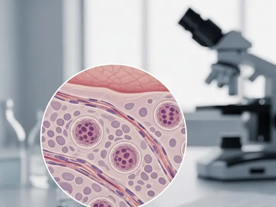
Clark Level II melanoma is a histological classification that describes the depth of invasion of melanoma cells within the skin. Developed by Dr. Wallace Clark, this system categorizes melanoma based on how far the cancerous cells have penetrated the different layers of the skin. Specifically, Clark Level II indicates that the melanoma cells have invaded the papillary dermis, which is the uppermost layer of the dermis, but have not yet extended into the deeper reticular dermis. This level of invasion is considered relatively superficial compared to higher Clark levels.
The **Clark Level 2 skin cancer meaning** is significant because the depth of invasion is a critical prognostic factor for melanoma. A lower Clark level generally suggests a less aggressive tumor with a lower risk of metastasis. For **understanding Clark Level 2 skin cancer**, it’s important to recognize that it represents an early stage of invasion. While the tumor has moved beyond the epidermis (the outermost layer of the skin), its confinement to the papillary dermis implies that it has not yet reached the deeper structures where blood vessels and lymphatic channels are more abundant, which could facilitate wider spread.
Pathologists determine the Clark level by examining a biopsy sample under a microscope. This assessment, along with the Breslow thickness (which measures the actual vertical thickness of the tumor in millimeters), provides a comprehensive picture of the melanoma’s invasiveness. According to the American Cancer Society, early detection of melanoma, when it is still superficial, significantly improves survival rates. For localized melanoma, the 5-year survival rate is very high, emphasizing the importance of classifications like Clark Level II in guiding timely intervention.
Key characteristics observed in a Clark Level II melanoma include:
- Invasion into the papillary dermis.
- Absence of invasion into the reticular dermis.
- Often associated with thin melanomas (low Breslow thickness).
- Typically detected during routine skin examinations or when a suspicious lesion is biopsied.
Clark Level II Melanoma Prognosis and Management
The **Clark Level II melanoma prognosis** is generally considered favorable, especially when compared to melanomas that have invaded deeper into the skin (Clark Levels III, IV, or V). Since the tumor is confined to the papillary dermis, the likelihood of regional or distant metastasis is lower. However, it is crucial to understand that Clark level is just one factor among several that influence prognosis. The Breslow thickness, presence of ulceration, mitotic rate, and lymph node involvement are also vital in determining the overall outlook and staging of melanoma.
Management for Clark Level II melanoma primarily involves complete surgical excision of the lesion. This procedure aims to remove the melanoma along with a margin of healthy surrounding tissue to ensure all cancerous cells are eradicated. The size of the surgical margin depends on the Breslow thickness of the tumor. For thin melanomas, which often correspond to Clark Level II, smaller margins are typically sufficient. Following surgery, patients are usually advised to undergo regular follow-up examinations to monitor for recurrence or the development of new lesions.
While Clark Level II indicates a relatively early stage, ongoing vigilance is essential. Patients are educated on self-skin exams and the importance of protecting their skin from UV radiation. Regular dermatological check-ups are recommended to detect any potential changes early. According to the National Cancer Institute, the 5-year relative survival rate for localized melanoma (which includes most Clark Level II cases) is approximately 99%, highlighting the effectiveness of early diagnosis and treatment.