
Abdominal Ultrasound
Abdominal ultrasound is a non-invasive medical imaging technique that uses high-frequency sound waves to create real-time images of organs and structures within the abdomen. It is a widely used diagnostic tool due to its safety, accessibility, and effectiveness in visualizing soft tissues.
Key Takeaways
- Abdominal ultrasound uses sound waves to visualize organs like the liver, kidneys, gallbladder, and pancreas.
- It is a safe, non-invasive procedure used to diagnose various conditions, from gallstones to tumors.
- Preparation typically involves fasting for several hours and sometimes drinking water to fill the bladder.
- The procedure is painless and usually takes 30-60 minutes, providing immediate real-time imaging.
- It helps healthcare providers assess organ size, structure, and detect abnormalities without radiation exposure.
What is Abdominal Ultrasound?
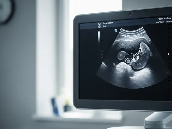
Abdominal Ultrasound is a diagnostic imaging method that employs sound waves to produce pictures of the internal organs located in the abdomen. This non-invasive technique uses a small transducer (probe) that emits sound waves and detects the echoes as they bounce off structures such as the liver, gallbladder, spleen, pancreas, kidneys, and major blood vessels. The echoes are then converted into real-time images displayed on a monitor, allowing healthcare professionals to assess the size, shape, and condition of these organs. The procedure is painless and does not involve radiation, making it a safe option for various patients, including pregnant women.
When considering an abdominal ultrasound procedure explained, it typically involves the patient lying on an examination table while a sonographer applies a special gel to the abdomen. This gel helps the transducer make full contact with the skin and eliminates air pockets that could interfere with the sound waves. The sonographer then moves the transducer across the skin, capturing images from different angles. Patients might be asked to hold their breath or change positions to improve image quality. The entire process usually takes between 30 to 60 minutes, and the images are reviewed by a radiologist who then prepares a report for the referring physician.
Uses and Benefits of Abdominal Ultrasound
The utility of abdominal ultrasound extends across a broad spectrum of medical diagnostics, offering significant advantages over other imaging modalities. The abdominal ultrasound uses and benefits are numerous, making it a cornerstone in the evaluation of many abdominal conditions.
Some primary uses include:
- Diagnosing Gallstones: Visualizing stones in the gallbladder and bile ducts, which can cause pain and inflammation.
- Assessing Liver Disease: Detecting conditions like fatty liver, cirrhosis, and liver masses.
- Evaluating Kidney Issues: Identifying kidney stones, cysts, tumors, and assessing blood flow to the kidneys.
- Investigating Pancreatic Disorders: Checking for inflammation (pancreatitis) or masses in the pancreas.
- Examining the Spleen: Assessing its size and detecting any abnormalities.
- Detecting Aortic Aneurysms: Screening for an enlargement of the main artery in the abdomen.
- Guiding Procedures: Assisting during biopsies or fluid drainage to ensure accuracy.
The benefits of this imaging technique are substantial. It is non-invasive, meaning it does not require needles, injections, or incisions. It uses no ionizing radiation, making it safe for repeated use and for sensitive populations like children and pregnant women. Furthermore, it provides real-time images, which allows for dynamic assessment of organ function and blood flow, and can be performed relatively quickly and is widely available.
Preparing for Your Abdominal Ultrasound
Proper preparation is crucial to ensure clear and accurate images during an abdominal ultrasound. Understanding how to prepare for abdominal ultrasound can significantly impact the success of the examination. Specific instructions will be provided by your healthcare provider or the imaging center, but general guidelines often include:
- Fasting: You will typically be asked to fast for 6 to 12 hours before the procedure. This means no food or drink (except plain water) to ensure your stomach and intestines are empty. Fasting is particularly important for visualizing the gallbladder, as food intake causes it to contract and empty, making it difficult to assess.
- Hydration (for Pelvic Organs): If your ultrasound includes the kidneys or bladder, you might be instructed to drink several glasses of water about an hour before your appointment and to avoid urinating. A full bladder helps to push bowel loops out of the way and provides a clear “window” for visualizing pelvic organs.
- Medications: Generally, you can continue to take your prescribed medications with a small sip of water, unless otherwise instructed by your doctor.
- Comfortable Clothing: Wear loose, comfortable clothing to your appointment, as you may need to expose your abdomen.
- Inform Your Provider: Always inform the imaging staff if you have any allergies, medical conditions, or are pregnant.
Following these instructions carefully helps the sonographer obtain the best possible images, leading to a more accurate diagnosis.


