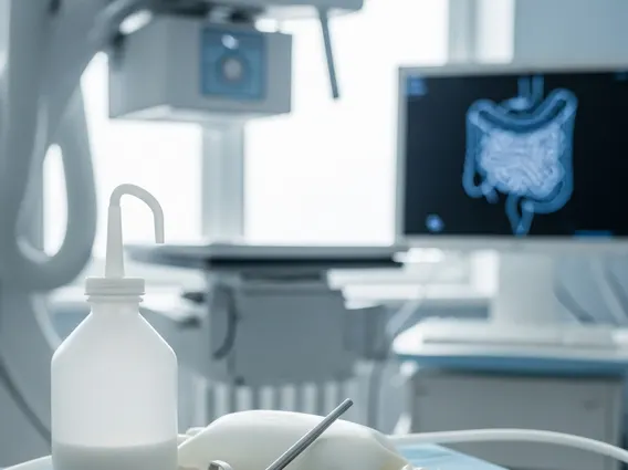
Double Balloon Enteroscopy
Double Balloon Enteroscopy (DBE) is an advanced endoscopic procedure designed to visualize and treat conditions within the small intestine, an area traditionally difficult to access. This technique allows for a thorough examination of the entire small bowel, offering both diagnostic insights and therapeutic interventions.
Key Takeaways
- Double Balloon Enteroscopy (DBE) is an advanced endoscopic technique for examining the small intestine.
- It uses two balloons to navigate and stabilize the endoscope, allowing deep access into the small bowel.
- The procedure can be performed antegrade (mouth) or retrograde (anus) depending on the area of interest.
- DBE is crucial for diagnosing and treating conditions like obscure gastrointestinal bleeding, Crohn’s disease, and small bowel tumors.
- It offers both diagnostic capabilities (biopsy, visualization) and therapeutic interventions (polypectomy, hemostasis).
What is Double Balloon Enteroscopy (DBE)?
Double Balloon Enteroscopy, often abbreviated as DBE, is a specialized endoscopic procedure that enables gastroenterologists to examine the entire length of the small intestine. Unlike conventional endoscopies that can only reach the initial part of the small bowel (duodenum) or the very end (terminal ileum), DBE utilizes a unique two-balloon system to navigate and stabilize the endoscope deep into the convoluted small intestine. This innovative approach allows for comprehensive visualization and intervention in areas previously inaccessible without surgery.
The technique was first developed in Japan and has since become a vital tool in gastroenterology. It involves an endoscope fitted with an inflatable balloon and an overtube, which is also equipped with a balloon. By alternately inflating and deflating these balloons, the endoscope can be “pleated” through the small bowel, allowing for significant advancement and a detailed inspection of the mucosa. This capability is particularly important for conditions affecting the middle sections of the small intestine.
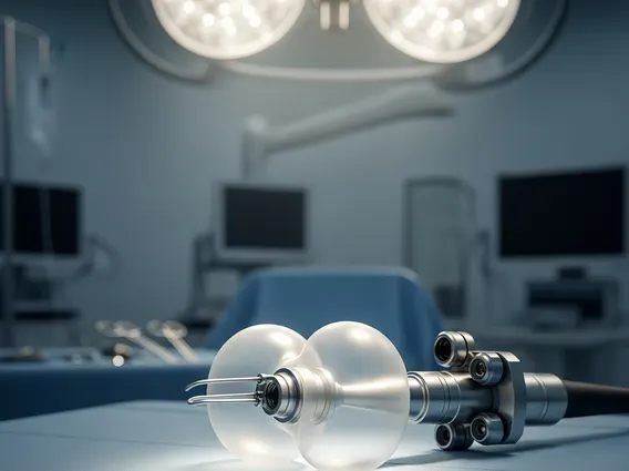
The Double Balloon Enteroscopy Procedure
The Double Balloon Enteroscopy procedure involves careful preparation and execution to ensure patient safety and diagnostic accuracy. Patients typically undergo a period of fasting, and bowel preparation may be required, especially for a retrograde approach. The procedure can be performed in one of two ways:
- Antegrade Approach: The endoscope is inserted through the mouth, passing through the esophagus, stomach, and duodenum, into the jejunum and ileum. This approach is generally preferred when the suspected pathology is in the upper or middle parts of the small intestine.
- Retrograde Approach: The endoscope is inserted through the anus, passing through the colon and into the ileum. This method is chosen when the suspected pathology is located in the distal ileum or when the antegrade approach has not reached the area of interest.
During the procedure, the patient is usually under conscious sedation or general anesthesia. The endoscope and overtube are advanced in a coordinated manner, with the balloons inflated and deflated to create a stable platform for advancement. This allows the physician to meticulously examine the small bowel lining, identify abnormalities, and perform necessary interventions. The duration of the procedure varies depending on the extent of examination required and any therapeutic interventions performed.
Diagnostic and Therapeutic Applications of DBE
The uses of Double Balloon Enteroscopy are extensive, encompassing both diagnostic and therapeutic applications for various small bowel conditions. Its ability to access the entire small intestine makes it invaluable for investigating symptoms that cannot be explained by conventional endoscopy or imaging techniques.
Key double balloon enteroscopy diagnostic uses include the investigation of obscure gastrointestinal bleeding, which accounts for a significant portion of small bowel examinations. DBE can pinpoint the source of bleeding, such as angioectasias, ulcers, or small tumors. It is also crucial for diagnosing and assessing the extent of Crohn’s disease in the small bowel, identifying strictures, fistulas, and inflammation. Furthermore, DBE is used to detect and characterize small bowel tumors, polyps, and other mucosal abnormalities that may be missed by other diagnostic modalities. Biopsies can be taken during the procedure to confirm diagnoses.
Beyond diagnosis, DBE offers significant therapeutic capabilities. Once a lesion is identified, the physician can perform various interventions through the endoscope. These include:
- Hemostasis (stopping bleeding) using clips, cautery, or injection therapy.
- Polypectomy (removal of polyps) to prevent potential malignancy.
- Dilation of strictures to improve patency and relieve symptoms.
- Foreign body retrieval from the small intestine.
- Placement of enteral feeding tubes beyond an obstruction.
According to a review published in the World Journal of Gastroenterology, DBE has a high diagnostic yield for obscure gastrointestinal bleeding, ranging from 50% to 75%, and allows for therapeutic intervention in a significant percentage of these cases. This highlights its critical role in managing complex small bowel disorders.