Cutaneous Leiomyoma
Cutaneous Leiomyoma refers to benign tumors that originate from the smooth muscle cells of the skin. While generally harmless, these lesions can cause significant discomfort and pain, impacting a patient’s quality of life.
Key Takeaways
- Cutaneous leiomyomas are benign smooth muscle tumors of the skin, often arising from arrector pili muscles.
- The primary symptom is pain, which can be spontaneous or triggered by cold, pressure, or touch.
- Diagnosis typically involves clinical examination followed by a skin biopsy for histological confirmation.
- Treatment options range from surgical excision for symptomatic lesions to various medical therapies aimed at pain management.
- These tumors can occur sporadically or be associated with hereditary conditions like hereditary leiomyomatosis and renal cell carcinoma (HLRCC).
What is Cutaneous Leiomyoma?
Cutaneous Leiomyoma is a type of benign tumor that develops from the smooth muscle tissue found within the skin. These tumors most commonly arise from the arrector pili muscles, which are small muscles attached to hair follicles responsible for making hair stand on end (goosebumps). Less frequently, they can originate from the smooth muscle of blood vessel walls or genital smooth muscle. While they are non-cancerous, their presence often leads to significant symptoms, primarily pain.
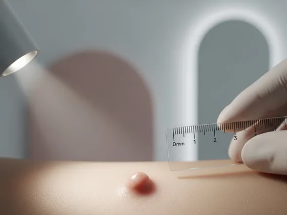
These lesions typically present as firm, reddish-brown to skin-colored nodules or papules, varying in size from a few millimeters to several centimeters. They can appear as solitary lesions but often occur as multiple tumors, sometimes clustered together. The exact prevalence of cutaneous leiomyomas is not well-documented due to their relatively uncommon nature, but they are recognized as a distinct dermatological condition that can significantly affect individuals due to associated pain.
Cutaneous Leiomyoma Symptoms, Causes, and Diagnosis
Understanding cutaneous leiomyoma symptoms causes is crucial for effective management. The hallmark symptom of cutaneous leiomyomas is pain, which can be spontaneous, paroxysmal (sudden and intense), or triggered by external stimuli such as cold temperatures, light touch, or pressure. This pain is often described as sharp, stabbing, or burning and can be quite severe, leading to considerable distress. The exact mechanism of pain is thought to involve nerve compression or muscle spasm within the tumor.
The causes of cutaneous leiomyomas can be sporadic or hereditary. Sporadic cases occur without a clear genetic link. However, multiple cutaneous leiomyomas can be a sign of hereditary leiomyomatosis and renal cell carcinoma (HLRCC), also known as Reed’s syndrome. This is an autosomal dominant genetic disorder caused by mutations in the fumarate hydratase (FH) gene, which also increases the risk of developing uterine leiomyomas (fibroids) and aggressive renal cell carcinoma. Therefore, individuals with multiple cutaneous leiomyomas should be evaluated for this underlying genetic condition.
Diagnosing cutaneous leiomyoma typically begins with a thorough clinical examination, where the physician assesses the appearance and tenderness of the skin lesions. However, a definitive diagnosis requires a skin biopsy. During this procedure, a small tissue sample is removed and examined under a microscope by a pathologist. Histopathological examination reveals characteristic bundles of smooth muscle cells arranged in an unorganized pattern, confirming the diagnosis. Immunohistochemical staining for smooth muscle actin (SMA) can further aid in confirmation.
- Clinical Presentation: Firm, skin-colored to reddish-brown nodules or papules.
- Pain Characteristics: Spontaneous, paroxysmal, or triggered by cold, pressure, or touch.
- Biopsy: Essential for definitive diagnosis, showing smooth muscle bundles.
- Genetic Testing: Recommended for multiple lesions to rule out HLRCC syndrome.
Cutaneous Leiomyoma Treatment Options
Managing cutaneous leiomyoma treatment options primarily focuses on alleviating pain and, in some cases, removing the lesions. Surgical excision is often the most effective treatment for symptomatic solitary or localized lesions, providing complete removal of the tumor and often leading to pain resolution. However, for multiple or widespread lesions, surgery may not be practical or may result in significant scarring, prompting the consideration of alternative therapies.
For patients with widespread or recurrent lesions, or those who are not candidates for surgery, various medical treatments aim to manage pain. These include oral medications such as calcium channel blockers (e.g., nifedipine), alpha-adrenergic blockers (e.g., phenoxybenzamine), and gabapentin, which can help reduce muscle spasms and neuropathic pain. Topical treatments, including local anesthetics or capsaicin cream, may also offer some relief. Additionally, botulinum toxin injections have shown promise in reducing pain by relaxing the smooth muscle within the tumors.
Other treatment modalities include cryotherapy, electrodessication, and laser ablation (e.g., CO2 laser), which can be used to destroy the lesions, particularly smaller ones. However, these methods may not always prevent recurrence or fully alleviate pain. It is important for patients to discuss all available options with their healthcare provider to determine the most appropriate and personalized treatment plan. Information on alternative or complementary therapies is supportive only and does not replace medical treatment.