Cin 1
Cervical Intraepithelial Neoplasia Grade 1 (CIN 1) represents a mild abnormality in the cells on the surface of the cervix, often detected during routine gynecological screenings. It is considered a low-grade precancerous condition, meaning it is not cancer but has the potential to progress if left unmonitored.

Key Takeaways
- CIN 1 is the mildest form of cervical intraepithelial neoplasia, indicating minor cellular changes on the cervix.
- The primary cause of CIN 1 is persistent infection with high-risk types of the human papillomavirus (HPV).
- Most cases of CIN 1 are asymptomatic and are typically discovered through routine Pap tests and subsequent colposcopy.
- A significant percentage of CIN 1 lesions spontaneously regress without intervention, particularly in younger individuals.
- Management often involves watchful waiting with regular follow-up, though treatment options like LEEP may be considered if the condition persists or progresses.
What is CIN 1 (Cervical Intraepithelial Neoplasia Grade 1)?
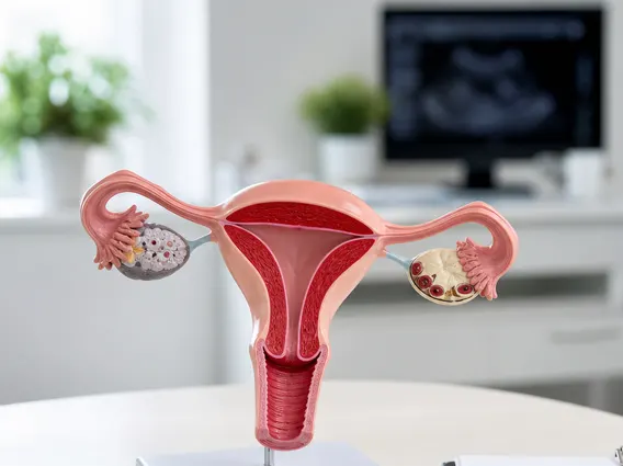
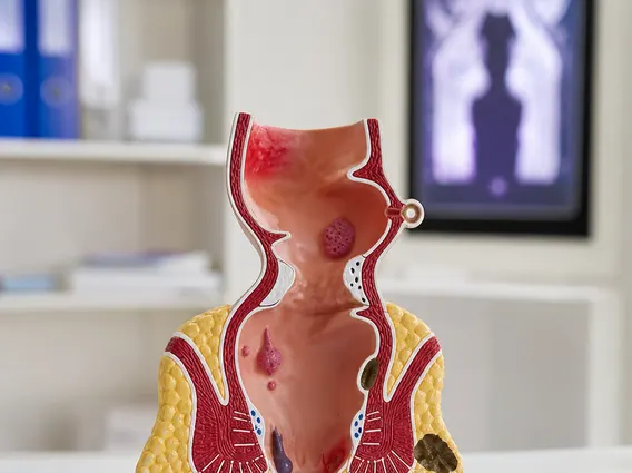
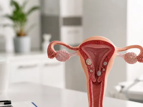
Cervical Intraepithelial Neoplasia Grade 1 (CIN 1) refers to a condition characterized by mild, abnormal cell changes on the surface of the cervix. These changes are not cancerous but are considered precancerous, indicating a low risk of progressing to cervical cancer. It is the least severe form of cervical intraepithelial neoplasia, affecting only the superficial layers of the cervical lining.
The presence of CIN 1 signifies that some cells in the cervical epithelium have started to grow abnormally. This condition is primarily linked to infection with high-risk types of the human papillomavirus (HPV), a very common sexually transmitted infection. While the term “neoplasia” might sound alarming, CIN 1 often resolves on its own as the body’s immune system clears the HPV infection and the abnormal cells return to normal.
Understanding CIN 1 Causes, Symptoms, and Diagnosis
The primary underlying factor for CIN 1 is persistent infection with high-risk strains of the human papillomavirus (HPV). HPV is a common virus, and while many infections clear naturally, some persistent infections can lead to cellular changes in the cervix. Other risk factors that may increase the likelihood of developing CIN 1 or its persistence include a weakened immune system, smoking, early age at first sexual intercourse, and multiple sexual partners. However, it’s crucial to understand that having these risk factors does not guarantee the development of CIN 1.
CIN 1 typically presents with no noticeable symptoms. Because it is asymptomatic, it is almost exclusively detected during routine cervical cancer screening tests. This underscores the importance of regular gynecological check-ups and Pap tests for early detection of any cervical abnormalities.
The diagnostic process for CIN 1 usually involves several steps:
- Pap Test (Pap Smear): This initial screening test collects cells from the cervix to check for abnormalities. An abnormal Pap test result, often indicating atypical squamous cells of undetermined significance (ASCUS) or low-grade squamous intraepithelial lesion (LSIL), frequently prompts further investigation.
- HPV Test: Often performed alongside or after an abnormal Pap test, this test identifies the presence of high-risk HPV types.
- Colposcopy: If a Pap test or HPV test is abnormal, a colposcopy is performed. This procedure uses a magnifying instrument to allow the clinician to visually examine the cervix for abnormal areas.
- Biopsy: During colposcopy, small tissue samples (biopsies) are taken from any suspicious areas. These samples are then examined under a microscope by a pathologist to confirm the diagnosis of CIN 1 and rule out higher-grade lesions or cancer.
CIN 1 Treatment Options and Management
The management of CIN 1 is often conservative due to its high rate of spontaneous regression. Many cases, especially in younger individuals, resolve without any intervention as the body’s immune system clears the HPV infection. According to the American Society for Colposcopy and Cervical Pathology (ASCCP) guidelines, active surveillance or watchful waiting is a common and appropriate approach for CIN 1.
Active surveillance involves regular follow-up examinations, typically including repeat Pap tests and/or HPV tests, and sometimes repeat colposcopies, at specified intervals (e.g., every 6-12 months). The goal is to monitor the lesion for regression or progression. If the CIN 1 persists for an extended period (e.g., two years) or shows signs of progression to a higher grade, treatment may then be considered.
When treatment for CIN 1 is deemed necessary, the primary goal is to remove or destroy the abnormal cells while preserving the healthy cervical tissue. Common treatment options include:
| Treatment Option | Description |
|---|---|
| Loop Electrosurgical Excision Procedure (LEEP) | A thin wire loop, heated by electricity, is used to remove the abnormal tissue from the cervix. This is a common and effective outpatient procedure. |
| Cryotherapy | Abnormal cells are frozen and destroyed using a very cold probe. This method is less commonly used for CIN 1 but may be an option in specific cases. |
| Laser Ablation | A laser beam is used to vaporize the abnormal cells on the surface of the cervix. |
After any treatment, continued follow-up is essential to ensure that all abnormal cells have been removed and to monitor for recurrence. It’s important to discuss the most suitable management plan with a healthcare provider, considering individual circumstances and risk factors.