Cat Scan
A Cat Scan, also known as a Computed Tomography (CT) scan, is a sophisticated medical imaging technique that plays a crucial role in diagnosing and monitoring various health conditions. This non-invasive procedure provides detailed cross-sectional images of the body, aiding healthcare professionals in making informed decisions.
Key Takeaways
- A Cat Scan (CT scan) uses X-rays and computer processing to create detailed cross-sectional images of the body.
- It works by rotating an X-ray source around the patient, capturing multiple images that are then reconstructed into 2D slices and 3D models.
- CT scans are widely used for diagnosing injuries, diseases, and guiding medical procedures across various body parts.
- Potential risks include exposure to ionizing radiation and possible reactions to contrast agents used in some procedures.
- The benefits of accurate diagnosis often outweigh the minimal risks associated with a single Cat Scan.
What is a Cat Scan (CT Scan) and How It Works
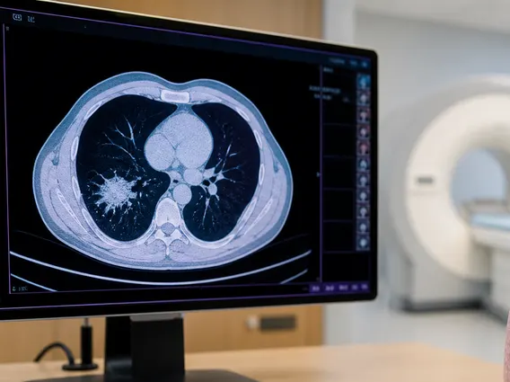
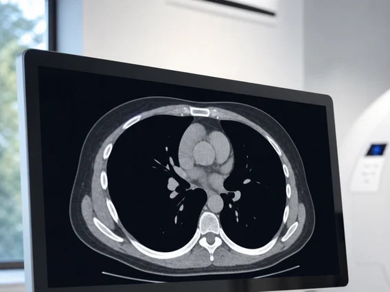
A Cat Scan, formally known as a Computed Tomography (CT) scan, refers to a diagnostic imaging procedure that combines a series of X-ray images taken from different angles around your body and uses computer processing to create cross-sectional images, or slices, of the bones, blood vessels, and soft tissues inside your body. These images provide much more detailed information than plain X-rays do.
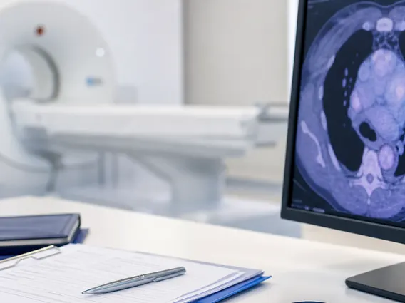
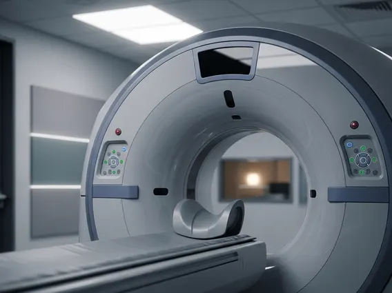
The process of how does a cat scan work involves a patient lying on a motorized table that slides into a large, donut-shaped machine called a gantry. Inside the gantry, an X-ray tube rotates around the patient, emitting a narrow beam of X-rays. On the opposite side of the patient, a detector array captures the X-rays that pass through the body. As the X-ray tube and detectors rotate, they collect hundreds of thousands of individual X-ray measurements from various angles. A powerful computer then processes these measurements to generate detailed cross-sectional images. These images can be viewed individually or stacked together to create a three-dimensional (3D) representation of the scanned area, allowing doctors to examine organs, bones, and tissues from multiple perspectives.
To enhance the visibility of specific structures, a contrast material may be administered orally, intravenously, or rectally. This material temporarily changes the way X-rays interact with certain tissues, making blood vessels, organs, or tumors appear brighter on the images. For instance, according to the American College of Radiology, contrast agents are used in approximately 30-50% of all CT scans to improve diagnostic accuracy.
Uses and Potential Risks of Cat Scan Procedures
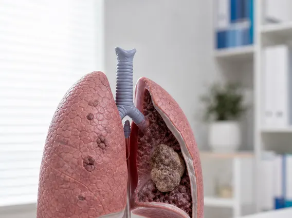
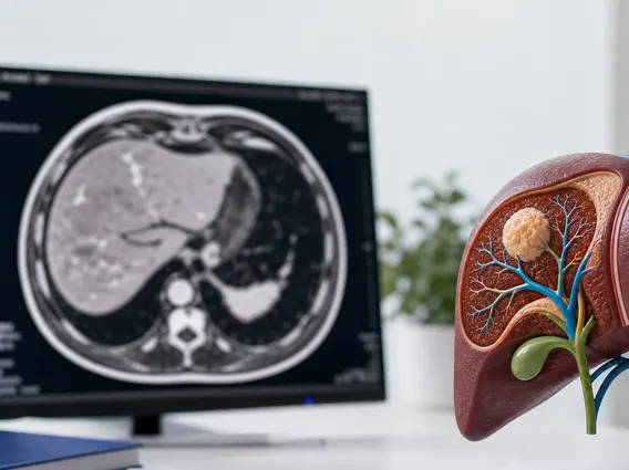
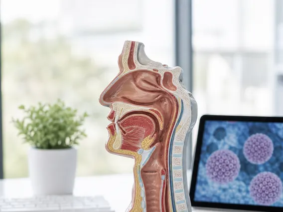
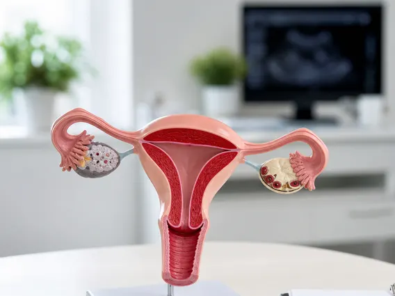
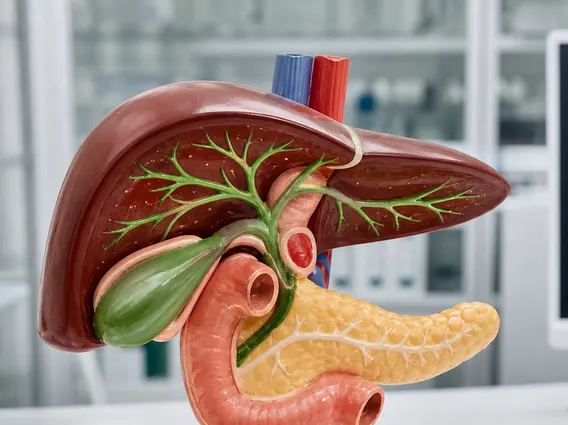
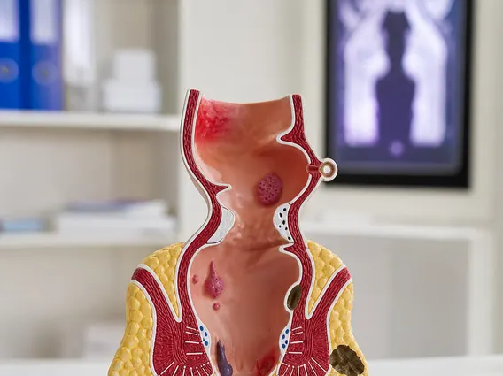
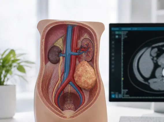
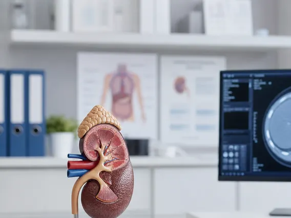
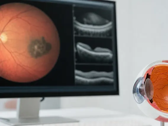
What are Cat Scans used for encompasses a broad range of medical applications, making them an indispensable tool in modern medicine. They are frequently employed to diagnose bone and joint problems, such as complex fractures and tumors, and to detect internal injuries and bleeding, particularly after trauma. CT scans are also vital for identifying diseases like cancer, heart disease, emphysema, and liver masses. Furthermore, they can guide procedures such as biopsies, surgery, and radiation therapy, helping physicians accurately target specific areas within the body. Common areas examined include the head (for stroke, tumors, or injury), chest (for lung conditions or heart issues), abdomen and pelvis (for digestive problems, kidney stones, or appendicitis), and spine (for herniated discs or spinal cord injuries).
While highly beneficial, there are certain risks of Cat Scan procedure that patients should be aware of. The primary concern is exposure to ionizing radiation. Although the amount of radiation from a single CT scan is relatively low, repeated exposure over time can slightly increase the lifetime risk of developing cancer. Healthcare providers carefully weigh the diagnostic benefits against this minimal risk, especially for children and pregnant women. According to the U.S. Food and Drug Administration (FDA), the typical radiation dose from a single diagnostic CT scan is generally considered safe, but unnecessary scans should be avoided.
Another potential risk involves allergic reactions to the contrast material, if used. These reactions can range from mild, such as hives or itching, to more severe, including difficulty breathing or anaphylaxis. Patients with a history of allergies, kidney disease, or diabetes are at a higher risk for adverse reactions and should inform their doctor before the procedure. Additionally, for individuals with kidney impairment, contrast agents can sometimes worsen kidney function. Patients are typically advised to drink plenty of fluids after a contrast-enhanced CT scan to help flush the material from their system.