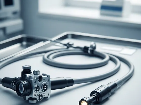
Gastroscope
A Gastroscope is a vital medical instrument used to examine the upper part of the gastrointestinal (GI) tract. This procedure allows healthcare professionals to visualize and assess the esophagus, stomach, and duodenum for various conditions.
Key Takeaways
- A Gastroscope is a flexible tube with a camera, used to visualize the upper GI tract.
- It helps diagnose conditions like ulcers, inflammation, and tumors, and can also be used for therapeutic interventions.
- The procedure involves careful insertion of the scope after sedation, allowing for direct examination and potential biopsies.
- Preparation typically includes fasting to ensure a clear view of the digestive organs.
- The examination provides detailed insights into the health of the esophagus, stomach, and duodenum.
What is a Gastroscope?
A Gastroscope is a long, thin, flexible tube equipped with a light source and a tiny camera at its tip. This advanced gastroscope medical device explanation highlights its role in allowing doctors to visually inspect the lining of the upper gastrointestinal tract, which includes the esophagus, stomach, and the first part of the small intestine (duodenum). The images captured by the camera are transmitted to a monitor, providing a real-time, magnified view of the internal organs.
Beyond simple visualization, modern gastroscopes often incorporate channels through which small instruments can be passed. These instruments enable various procedures, such as taking tissue samples (biopsies), removing polyps, or controlling bleeding. The flexibility of the scope allows it to navigate the natural curves of the digestive system, minimizing discomfort while maximizing diagnostic and therapeutic capabilities.
What is a Gastroscope Used For?
A gastroscope is primarily used for both diagnostic and therapeutic purposes within the upper GI tract. Diagnostically, it helps identify the causes of symptoms such as persistent heartburn, difficulty swallowing, abdominal pain, nausea, vomiting, or unexplained weight loss. It can detect conditions including:
- Esophagitis (inflammation of the esophagus)
- Gastritis (inflammation of the stomach lining)
- Peptic ulcers (sores in the stomach or duodenum)
- Celiac disease (damage to the small intestine from gluten)
- Barrett’s esophagus (a precancerous condition)
- Tumors or cancerous growths
Therapeutically, a gastroscope enables physicians to perform minor interventions directly during the examination. For instance, biopsies can be taken to test for Helicobacter pylori infection or to analyze suspicious tissue. Polyps, which are abnormal growths, can be removed to prevent them from becoming cancerous. The scope can also be used to stop active bleeding from ulcers or varices, dilate narrowed areas (strictures), or retrieve foreign objects that have been swallowed. According to the American Society for Gastrointestinal Endoscopy (ASGE), upper endoscopy, which utilizes a gastroscope, is one of the most common and effective procedures for evaluating and treating upper GI conditions, with millions performed annually worldwide.
How Does a Gastroscope Procedure Work?
The gastroscope procedure, also known as an upper endoscopy, typically begins with patient preparation. Patients are usually asked to fast for several hours before the procedure to ensure the stomach is empty, allowing for a clear view and reducing the risk of aspiration. Upon arrival, a local anesthetic spray may be applied to the back of the throat to numb it, and intravenous (IV) sedation is often administered to help the patient relax and minimize discomfort during the examination. This sedation usually induces a drowsy state, and some patients may even fall asleep.
Once the patient is sedated, they are positioned on their side, and a mouth guard is placed to protect their teeth and the scope. The physician then gently inserts the gastroscope into the mouth, guiding it down the esophagus, through the stomach, and into the duodenum. Air is often inflated through the scope to gently expand the GI tract, providing a better view of the lining. The physician carefully examines the walls of these organs on a monitor, looking for any abnormalities. During this process, the gastroscope examination details, such as the color of the tissue, presence of lesions, or any bleeding, are meticulously observed and recorded. If necessary, small instruments are passed through the scope to perform biopsies or other therapeutic interventions. The entire procedure typically takes 15 to 30 minutes. After the examination, the scope is slowly withdrawn, and the patient is monitored in a recovery area until the effects of the sedation wear off. Patients are advised against driving or operating machinery for the rest of the day due to the lingering effects of sedation.