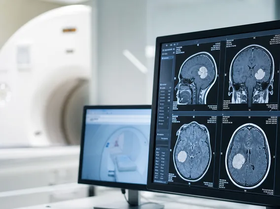
Functional Magnetic Resonance Imaging
Functional Magnetic Resonance Imaging (fMRI) is a specialized neuroimaging technique that measures brain activity by detecting changes associated with blood flow. This non-invasive method provides crucial insights into how different parts of the brain respond during various tasks or states.

Key Takeaways
- Functional Magnetic Resonance Imaging (fMRI) is a non-invasive technique that measures brain activity by detecting changes in blood flow.
- It relies on the Blood-Oxygen-Level Dependent (BOLD) signal, which differentiates between oxygenated and deoxygenated blood.
- fMRI is widely used in neuroscience for brain mapping, understanding cognitive processes, and pre-surgical planning.
- Benefits include its non-invasive nature and lack of radiation, while risks primarily involve magnetic field interactions and claustrophobia.
What is Functional Magnetic Resonance Imaging (fMRI)?
Functional Magnetic Resonance Imaging (fMRI) is a sophisticated neuroimaging technique that observes brain activity by measuring changes in blood flow. When a specific area of the brain becomes active, it demands more oxygen and nutrients, leading to an increase in localized blood flow. fMRI detects these subtle hemodynamic changes, allowing researchers and clinicians to map which parts of the brain are involved in particular mental processes, such as thinking, feeling, moving, or perceiving.
Unlike traditional MRI, which provides detailed anatomical images of the brain, fMRI focuses on dynamic physiological changes. It offers a unique window into the brain’s functional organization, making it an invaluable tool for understanding both healthy brain function and the impact of neurological conditions. This non-invasive method does not involve ionizing radiation, making it a safer option for repeated studies compared to techniques like PET scans.
How Functional Magnetic Resonance Imaging Works
The core principle behind how functional magnetic resonance imaging works lies in the detection of the Blood-Oxygen-Level Dependent (BOLD) signal. When neurons in a specific brain region become active, they consume oxygen. To replenish this oxygen, the body increases blood flow to that area, delivering more oxygenated blood than is immediately used. Hemoglobin, the protein in red blood cells that carries oxygen, has different magnetic properties depending on whether it is oxygenated or deoxygenated.
Oxygenated hemoglobin is diamagnetic, meaning it is weakly repelled by a magnetic field, while deoxygenated hemoglobin is paramagnetic, meaning it is weakly attracted to a magnetic field. An fMRI scanner uses a powerful magnetic field and radio waves to detect these differences. Areas with increased neural activity show a higher ratio of oxygenated to deoxygenated blood, which results in a stronger fMRI signal. The scanner then processes these signals to create detailed maps of brain activity, highlighting regions that are more active during specific tasks or stimuli.
Applications and Safety of fMRI
Functional Magnetic Resonance Imaging has revolutionized the field of neuroscience, offering unprecedented insights into brain function. The primary uses of fMRI in neuroscience span a wide range of research and clinical applications. It is extensively used to map brain regions responsible for various cognitive functions, such as language processing, memory recall, emotional responses, and motor control. This capability is crucial for understanding the neural basis of human behavior and cognition.
Clinically, fMRI plays a vital role in pre-surgical planning for patients undergoing brain surgery, particularly for tumor removal or epilepsy treatment. By identifying critical areas like those controlling speech or movement, surgeons can minimize damage to essential brain functions. Furthermore, fMRI is a powerful tool for studying neurological and psychiatric disorders, helping researchers understand the functional changes associated with conditions like Alzheimer’s disease, depression, and schizophrenia. For instance, fMRI can reveal altered connectivity patterns or reduced activity in specific brain networks.
Regarding the benefits and risks of fMRI scan, it is generally considered a safe procedure. Its non-invasive nature and the absence of ionizing radiation are significant advantages, making it suitable for pediatric patients and repeated studies. However, certain considerations and potential risks exist, primarily due to the strong magnetic field involved.
| Benefits of fMRI Scan | Risks of fMRI Scan |
|---|---|
| Non-invasive, no ionizing radiation exposure. | Strong magnetic field poses risks for patients with metallic implants (e.g., pacemakers, certain surgical clips). |
| Provides high-resolution images of brain activity. | Loud noise from the scanner can be uncomfortable; hearing protection is provided. |
| Helps localize brain functions for research and surgical planning. | Claustrophobia can be an issue for some individuals due to the enclosed space. |
| Can detect subtle changes in blood flow related to neural activity. | Potential for minor skin burns from contact with scanner parts or wires, though rare. |
Patients are thoroughly screened for any metallic implants or conditions that might contraindicate an fMRI scan. Overall, the diagnostic and research benefits of fMRI typically outweigh its minimal risks, especially when performed under strict safety protocols.