Fluoroscopy
Fluoroscopy is a medical imaging technique that uses X-rays to obtain real-time, moving images of the internal structures of a patient. Often described as an “X-ray movie,” this dynamic imaging method allows clinicians to visualize the motion of organs and the flow of contrast agents through the body.
Key Takeaways
- Fluoroscopy provides real-time X-ray images, enabling dynamic visualization of internal body processes.
- It works by passing a continuous X-ray beam through the body, with images displayed on a monitor.
- Contrast agents are frequently used to highlight specific organs or vessels during the procedure.
- Common applications include guiding catheter placements, evaluating digestive system function, and assessing joint movement.
- The primary benefit is its ability to show function and movement, which static X-rays cannot provide.
What is Fluoroscopy?
Fluoroscopy refers to a specialized medical imaging technique that produces continuous X-ray images, much like a live video feed. Unlike traditional X-rays, which capture static snapshots, fluoroscopy allows healthcare professionals to observe internal body structures in motion. This dynamic capability is crucial for diagnosing various conditions and guiding intricate medical procedures. The process involves a continuous X-ray beam passing through the body, with the resulting images displayed on a monitor in real-time.
What is fluoroscopy imaging? It is essentially a diagnostic tool that provides a live view of internal organs, bones, and tissues. This real-time visualization is particularly valuable when assessing the function of organ systems, such as the digestive tract, or when precise guidance is needed during minimally invasive surgeries. Often, a contrast agent, such as barium or iodine, is introduced into the body to enhance the visibility of specific structures, making them stand out against surrounding tissues.
How Does Fluoroscopy Work?
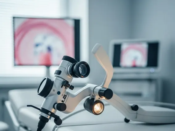
How does fluoroscopy work? The fundamental principle involves a continuous X-ray beam directed through the patient’s body. On the opposite side of the patient, an image intensifier or a flat-panel detector captures the X-rays that pass through the body. These captured X-rays are then converted into visible light images, which are immediately displayed on a high-resolution monitor. This continuous capture and display create a live, moving image, allowing medical professionals to observe physiological processes as they happen.
To enhance the visibility of specific organs or blood vessels, contrast agents are frequently administered. For instance, barium sulfate is commonly used to visualize the gastrointestinal tract, while iodine-based contrast is often injected into blood vessels to highlight arteries and veins. As the contrast agent moves through the body, its path and interaction with tissues become visible on the fluoroscopy monitor, providing critical diagnostic information. The ability to see these internal movements in real-time is what distinguishes fluoroscopy from other static imaging methods, offering a unique perspective on bodily functions and anatomical structures.
Uses and Benefits of Fluoroscopy
Fluoroscopy offers a wide range of diagnostic and interventional applications across various medical specialties. Its primary benefit lies in its ability to provide dynamic, real-time visualization, which is indispensable for procedures requiring precise guidance and for evaluating the functional aspects of internal organs. This capability allows clinicians to make immediate adjustments during procedures and accurately assess the movement and interaction of bodily systems.
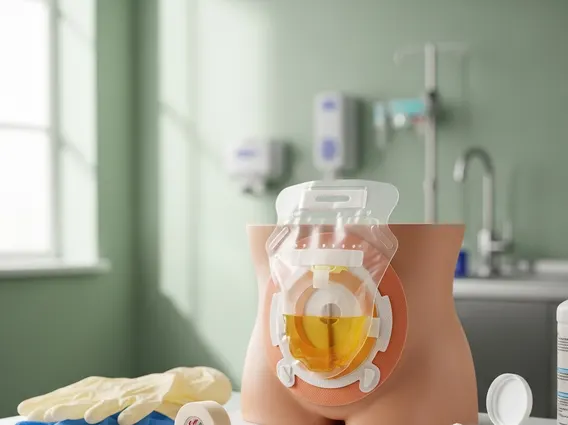
The Fluoroscopy procedure explanation often highlights its role in guiding complex interventions. For example, it is routinely used to guide the placement of catheters, stents, and other devices during cardiac procedures, vascular interventions, and pain management injections. In gastroenterology, it helps diagnose swallowing disorders, assess the movement of food through the digestive tract (e.g., barium swallow, upper GI series), and identify blockages or abnormalities. Orthopedic surgeons use fluoroscopy to guide joint injections and assist in fracture reductions. The benefits extend to:
- Precise Guidance: Enables accurate placement of medical instruments during minimally invasive procedures, reducing risks and improving outcomes.
- Dynamic Assessment: Allows real-time observation of organ function, such as heart contractions, blood flow, or digestive motility.
- Immediate Feedback: Clinicians can instantly see the effects of interventions and make necessary adjustments.
- Versatility: Applicable across numerous medical fields, from cardiology and gastroenterology to orthopedics and urology.
These applications underscore why fluoroscopy uses and benefits are so significant in modern medicine, providing essential insights that static imaging cannot offer.