Film Mammography
Film Mammography is a traditional medical imaging technique used to examine breast tissue for the early detection and diagnosis of breast diseases, primarily breast cancer. It plays a crucial role in women’s health by providing detailed images that can reveal abnormalities before they are palpable.
Key Takeaways
- Film Mammography is a conventional X-ray method for breast imaging, vital for breast cancer screening.
- The process involves breast compression and X-ray exposure, with images recorded on specialized film.
- It has a long history of effectiveness but involves physical film development and storage.
- While offering benefits like established efficacy, it presents risks such as radiation exposure and potential discomfort.
- Digital mammography has largely superseded film, offering advantages in image quality, storage, and dose management.
What is Film Mammography?
Film Mammography refers to a specialized type of X-ray imaging used to create detailed pictures of the breast. For decades, it has been the standard screening tool for the early detection of breast cancer and other breast conditions. This diagnostic procedure helps identify abnormalities, such as tumors, cysts, or microcalcifications, which may be too small to be felt during a physical examination. Early detection through mammography significantly improves treatment outcomes and survival rates for breast cancer patients. According to the World Health Organization (WHO), breast cancer is the most common cancer among women globally, underscoring the importance of effective screening methods like mammography.
How Film Mammography Works
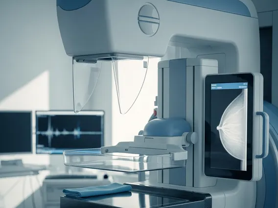
The process of Film Mammography involves several key steps designed to produce clear images of breast tissue. During the procedure, the patient’s breast is carefully positioned on a specialized X-ray plate and gently compressed between two paddles. This compression is essential for several reasons: it flattens the breast tissue to ensure all layers are visible, reduces the amount of radiation needed, minimizes motion blur, and separates overlapping tissues to enhance image clarity. Once compressed, a low-dose X-ray beam passes through the breast tissue and strikes a film cassette located beneath it. This cassette contains a special film that is sensitive to X-rays. The X-rays that penetrate the breast tissue expose the film, creating a latent image. After exposure, the film cassette is removed and processed in a darkroom using chemical solutions, similar to traditional photography, to develop the image. The resulting image, known as a mammogram, is a physical film that radiologists then examine on a light box for any signs of abnormalities.
Film Mammography: Benefits, Risks, and Comparison to Digital
Film mammography benefits and risks are important considerations when evaluating this traditional imaging method. Historically, its primary benefit has been its proven effectiveness in reducing breast cancer mortality through early detection. It has been widely available and relatively cost-effective, making it accessible for broad screening programs. However, there are also associated risks. Patients experience a small amount of radiation exposure during the procedure, though modern equipment is designed to minimize this. Discomfort or pain from breast compression is common but temporary. Furthermore, film mammograms can sometimes produce false positives, leading to unnecessary biopsies, or false negatives, where a cancer is missed.
The landscape of breast imaging has significantly evolved with the advent of digital technology. The comparison of Film vs digital mammography highlights several key differences:
| Feature | Film Mammography | Digital Mammography |
|---|---|---|
| Image Acquisition | X-rays expose a physical film cassette. | X-rays are converted into electrical signals by detectors. |
| Image Storage | Physical films requiring dedicated storage. | Digital files stored electronically (PACS). |
| Image Quality | Fixed contrast and density; harder to adjust post-acquisition. | Adjustable contrast, brightness, and zoom post-acquisition; often clearer for dense breasts. |
| Radiation Dose | Generally slightly higher than digital, though still low. | Often uses a lower radiation dose, especially with advanced techniques. |
| Interpretation | Radiologists view physical films on a light box. | Radiologists view images on high-resolution computer monitors. |
| Workflow | Requires film processing, slower. | Instant image review, faster workflow. |
Digital mammography has largely replaced film mammography in many clinical settings due to its advantages in image manipulation, easier storage and retrieval, and often lower radiation dose. While film mammography remains a viable option in some areas, digital systems offer enhanced diagnostic capabilities, particularly for women with dense breast tissue, and facilitate easier sharing of images for second opinions.