Pecoma
Pecoma refers to a rare group of mesenchymal tumors that exhibit perivascular epithelioid cell differentiation. These tumors can arise in various organs throughout the body, though they are most commonly associated with the kidney, liver, and uterus.

Key Takeaways
- Pecoma is a rare tumor type characterized by perivascular epithelioid cell differentiation.
- It can affect multiple organs, with the kidney, liver, and uterus being common sites.
- Symptoms vary widely depending on the tumor’s location and size.
- Diagnosis typically involves imaging, biopsy, and immunohistochemical staining.
- Treatment often includes surgical removal, with targeted therapies considered for advanced cases.
What is Pecoma?
Pecoma is an acronym for Perivascular Epithelioid Cell Tumor, a distinctive and uncommon type of mesenchymal neoplasm. These tumors are characterized by their unique cellular composition, featuring epithelioid cells that often surround blood vessels. While Pecoma can occur sporadically, it is notably associated with tuberous sclerosis complex (TSC), a genetic disorder that predisposes individuals to the development of benign and malignant tumors in various organs. The exact prevalence of Pecoma is challenging to determine due to its rarity, but it is considered a significant clinical entity, particularly in the context of TSC. According to a review published in the journal Cancer Research, Pecoma represents a spectrum of lesions, ranging from benign to potentially malignant, with their biological behavior often difficult to predict based solely on histological features.
The causes of Pecoma are not fully understood, but a strong genetic link exists, especially with mutations in the TSC1 or TSC2 genes, which are responsible for encoding hamartin and tuberin proteins, respectively. These proteins act as tumor suppressors, and their dysfunction can lead to uncontrolled cell growth and proliferation, contributing to the development of Pecoma. While the majority of Pecoma cases are benign, a subset can exhibit aggressive features, including rapid growth, metastasis, and recurrence, necessitating careful monitoring and management.
Pecoma Symptoms and Diagnosis
The presentation of Pecoma symptoms and diagnosis is highly variable, largely depending on the tumor’s location, size, and whether it is exerting pressure on surrounding tissues or organs. Many Pecoma tumors are discovered incidentally during imaging studies performed for other conditions, as they may remain asymptomatic for extended periods. When symptoms do occur, they can include:
- Abdominal pain or discomfort: Common with tumors in the kidney, liver, or gastrointestinal tract.
- Palpable mass: A noticeable lump or swelling, particularly in superficial locations or when tumors grow large.
- Bleeding: Hemorrhage can occur, especially in uterine Pecoma, leading to abnormal vaginal bleeding.
- Systemic symptoms: In rare aggressive cases, patients might experience fatigue, weight loss, or fever.
Diagnosis typically begins with imaging studies such as ultrasound, computed tomography (CT) scans, or magnetic resonance imaging (MRI), which can help locate the tumor and assess its size and characteristics. However, definitive diagnosis requires a tissue biopsy, followed by histopathological examination and immunohistochemical staining. Pecoma cells typically stain positive for melanocytic markers (like HMB-45, Melan-A) and muscle markers (like smooth muscle actin), which helps distinguish them from other tumor types. This comprehensive diagnostic approach is crucial for accurate classification and guiding appropriate treatment strategies.
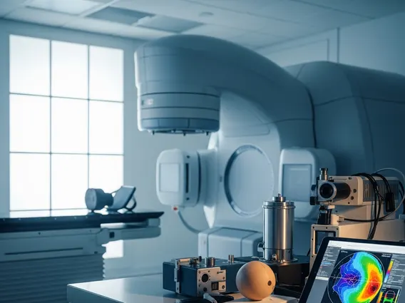
Pecoma Treatment Options
The selection of Pecoma treatment options is highly individualized, taking into account the tumor’s location, size, whether it is benign or malignant, and the patient’s overall health. For localized and resectable tumors, surgical excision is often the primary and most effective treatment. The goal of surgery is to remove the entire tumor with clear margins to prevent recurrence. For instance, in renal Pecoma, partial or radical nephrectomy may be performed, depending on the tumor’s size and involvement of the kidney.
In cases where the tumor is unresectable, metastatic, or shows aggressive features, other therapeutic approaches may be considered. Targeted therapies, particularly those inhibiting the mTOR (mammalian target of rapamycin) pathway, have shown promise. Drugs like everolimus and sirolimus, which are mTOR inhibitors, can be effective, especially for Pecoma associated with tuberous sclerosis complex, as the TSC1/TSC2 gene mutations directly impact this pathway. These therapies work by blocking signals that promote cell growth and division, thereby slowing or stopping tumor progression. While chemotherapy and radiation therapy are generally less effective for Pecoma, they may be considered in very specific, aggressive scenarios or as palliative measures. Regular follow-up and surveillance are essential after treatment to monitor for any signs of recurrence or new tumor development.