3 D Mammography
3D Mammography, also known as Digital Breast Tomosynthesis, represents a significant advancement in breast imaging technology, offering a more detailed view of breast tissue compared to traditional methods. This innovative screening tool plays a crucial role in the early detection and diagnosis of breast cancer, enhancing diagnostic accuracy for many individuals.
Key Takeaways
- 3D Mammography (Digital Breast Tomosynthesis) captures multiple X-ray images from different angles to create a three-dimensional reconstruction of the breast.
- This advanced imaging technique helps radiologists identify abnormalities more clearly, especially in dense breast tissue.
- It significantly improves cancer detection rates and reduces the number of false positives, leading to fewer unnecessary recalls for additional imaging.
- Compared to 2D mammography, 3D Mammography provides superior clarity and detail, aiding in earlier and more accurate diagnoses.
What is 3D Mammography (Digital Breast Tomosynthesis)?
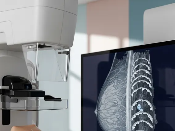
3D Mammography, or Digital Breast Tomosynthesis (DBT), is an advanced type of breast imaging that uses a low-dose X-ray system to create multiple images of the breast from different angles. These images are then reconstructed by a computer into a three-dimensional volume, allowing radiologists to view the breast tissue in thin, detailed layers. This layered approach helps overcome the limitations of conventional 2D mammography, where overlapping breast tissue can obscure abnormalities or create false alarms.
The primary goal of 3D Mammography is to enhance the detection of breast cancer at its earliest stages, particularly for individuals with dense breast tissue, which can be challenging to evaluate with standard mammograms. By providing a clearer, more comprehensive view, DBT improves diagnostic accuracy and contributes to more effective screening programs.
How Does 3D Mammography Work?
3D Mammography operates using a specialized X-ray machine that moves in an arc over the breast, capturing a series of low-dose images from various angles. During the procedure, the breast is compressed, similar to a traditional mammogram, to ensure consistent image quality and minimize radiation exposure. However, instead of a single static image, the tomosynthesis unit collects approximately 11 images in a sweep across the breast in just a few seconds.
Once these digital images are acquired, a computer processes them to generate a stack of thin, high-resolution “slices” of breast tissue. Radiologists can then scroll through these individual slices, much like flipping through pages of a book, to examine the breast tissue layer by layer. This ability to visualize tissue in cross-section helps differentiate between overlapping normal breast tissue and actual lesions, making it easier to detect small cancers and reducing the likelihood of misinterpreting benign findings.
Benefits of 3D Mammography and 2D Comparison
The benefits of 3D Mammography are substantial, particularly when compared to traditional 2D mammography. One of the most significant advantages is its improved ability to detect invasive breast cancer. Studies have shown that 3D Mammography can increase cancer detection rates by 15-30% compared to 2D mammography alone, especially in women with dense breasts. This enhanced detection leads to earlier diagnosis and potentially more favorable treatment outcomes.
Another key benefit is the reduction in recall rates. Overlapping tissue in 2D mammograms often leads to “false positives,” where a woman is called back for additional imaging or biopsies due to suspicious findings that turn out to be benign. 3D Mammography’s ability to separate tissue layers significantly reduces these false positives, decreasing patient anxiety and the need for unnecessary follow-up procedures. According to the American Cancer Society, 3D mammography has been shown to reduce recall rates by up to 15%.
To further illustrate the differences, here is a comparison of 3D Mammography vs 2D Mammography:
| Feature | 2D Mammography | 3D Mammography (Digital Breast Tomosynthesis) |
|---|---|---|
| Image Type | Two-dimensional flat images | Multiple images reconstructed into a 3D volume (thin slices) |
| Tissue Overlap | Significant, can obscure abnormalities | Minimized, allows clear visualization of layers |
| Cancer Detection | Standard detection rates | Higher detection rates, especially for invasive cancers |
| Recall Rates | Higher due to false positives | Lower, fewer unnecessary callbacks |
| Dense Breasts | More challenging to interpret | Improved visualization and detection |
| Radiation Dose | Low | Slightly higher than 2D, but within FDA guidelines and clinically acceptable |
The enhanced clarity and reduced ambiguity provided by 3D Mammography make it a valuable tool in breast cancer screening, offering peace of mind and more accurate results for many patients.