3 D
3 D refers to the three-dimensional representation and analysis of medical data, a revolutionary approach transforming diagnostics, treatment planning, and surgical procedures. This advanced methodology allows healthcare professionals to visualize complex anatomical structures with unprecedented depth and clarity.
Key Takeaways
- 3 D technology in medicine provides enhanced visualization of anatomical structures, improving diagnostic accuracy and treatment planning.
- The process involves acquiring multiple 2D images or volumetric data, which are then reconstructed into a comprehensive 3D model.
- Various types of 3D imaging, including CT, MRI, and ultrasound, offer distinct advantages for different clinical applications.
- The evolution of 3D imaging from early stereoscopes to modern digital reconstruction has significantly advanced medical capabilities.
What is 3 D Technology?
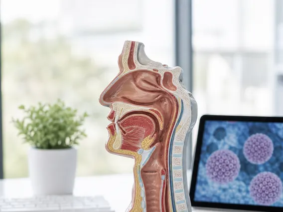
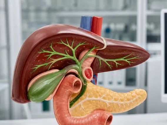
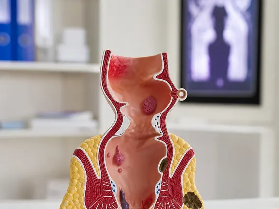
3 D refers to the medical application of three-dimensional imaging and visualization techniques, providing depth perception and spatial relationships that are crucial for understanding complex biological structures. This technology moves beyond traditional two-dimensional views, offering a comprehensive perspective essential for accurate diagnosis and precise intervention. What is 3d technology in this context? It encompasses a range of sophisticated tools and methods used to create detailed, volumetric representations of organs, tissues, and pathologies within the human body. These representations are invaluable for surgical planning, radiation therapy, and patient education.
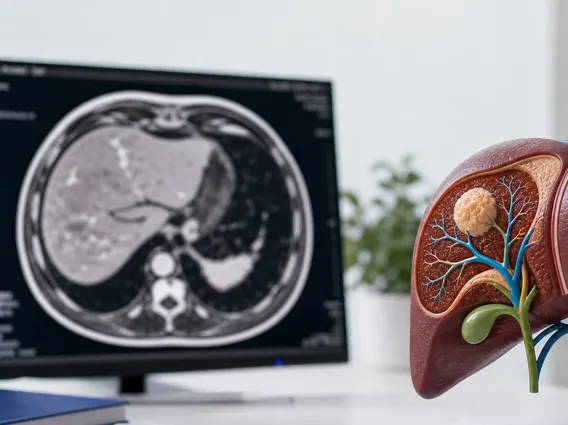
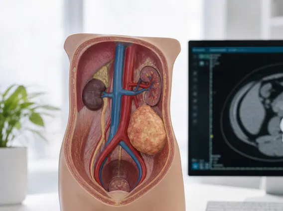
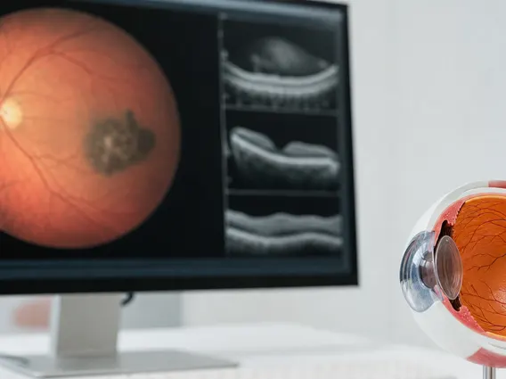
The integration of 3 D capabilities has profoundly impacted various medical fields. For instance, in oncology, it enables clinicians to precisely delineate tumor boundaries, assess their relationship to surrounding vital structures, and monitor treatment response more effectively. According to the World Health Organization (WHO), advanced imaging techniques are increasingly vital in the early detection and management of non-communicable diseases, including cancer, where 3 D visualization plays a pivotal role in improving outcomes. This enhanced spatial understanding aids in minimizing damage to healthy tissue during procedures, ultimately leading to safer and more effective patient care.
How 3D Imaging Works
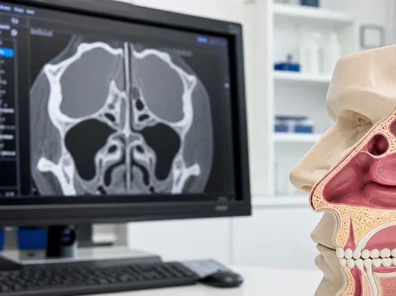
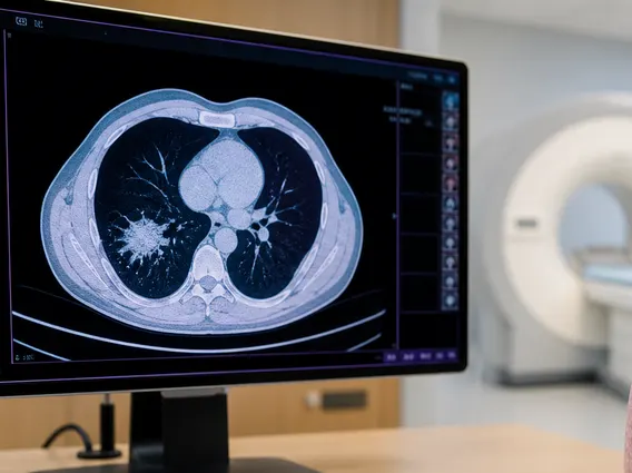
The fundamental principle behind how does 3d work explained in medical imaging involves acquiring data from multiple angles or slices and then computationally reconstructing these individual pieces into a cohesive three-dimensional model. This process typically begins with a data acquisition phase, where specialized scanners capture a series of two-dimensional images or volumetric data sets. For example, a Computed Tomography (CT) scanner rotates around the patient, taking numerous X-ray projections, while Magnetic Resonance Imaging (MRI) collects signals from atomic nuclei within the body at different spatial locations.
Once the raw data is collected, powerful computer algorithms process and combine these individual data points. This reconstruction involves complex mathematical transformations that convert the planar information into a volumetric dataset, allowing for interactive visualization from any angle. Advanced rendering techniques then create realistic images, often with color and texture mapping, to highlight specific anatomical features or pathological areas. This ability to manipulate and explore the 3D model virtually provides clinicians with an unparalleled view, helping them to identify abnormalities, measure dimensions, and plan interventions with greater precision.
Types of 3D and Its Historical Development
The field of medical imaging has seen the emergence of different types of 3d technologies, each leveraging distinct physical principles to generate three-dimensional data. Key modalities include:
- Computed Tomography (CT): Uses X-rays to create cross-sectional images, which are then stacked to form a 3D model, excellent for bone and soft tissue visualization.
- Magnetic Resonance Imaging (MRI): Employs strong magnetic fields and radio waves to produce detailed images of soft tissues, particularly useful for brain, spinal cord, and joint imaging.
- 3D Ultrasound: Utilizes sound waves to capture volumetric data in real-time, commonly used in obstetrics and cardiology for dynamic visualization.
- Positron Emission Tomography (PET) and Single-Photon Emission Computed Tomography (SPECT): Nuclear medicine techniques that provide functional 3D images, revealing metabolic activity or blood flow.
The history of 3d imaging traces back to early attempts at stereoscopic vision. While the concept of perceiving depth has existed for centuries, its application in medicine began to truly flourish with the advent of digital computing. Early pioneers in the 1970s and 1980s developed algorithms for reconstructing 3D images from CT and MRI scans. These initial systems were rudimentary but laid the groundwork for the sophisticated visualization tools available today. Significant milestones include the development of volume rendering techniques, which allowed for more realistic and interactive displays, and the integration of 3D data into surgical navigation systems. This continuous evolution has transformed medical practice, moving from static 2D images to dynamic, interactive 3D models that enhance diagnostic capabilities and guide complex procedures.