2 Dimensional Mammography
2 Dimensional Mammography is a foundational imaging technique used for breast cancer screening and diagnosis. This article explores the principles, advantages, and key differences compared to more advanced modalities.
Key Takeaways
- 2 Dimensional Mammography (2D mammography) is a standard X-ray imaging technique for breast cancer detection.
- It works by compressing the breast and taking two primary views, providing a flattened image.
- Advantages include widespread availability, lower cost, and established effectiveness in screening.
- While effective, it can have limitations in dense breast tissue due to tissue overlap.
- It differs from 3D mammography (tomosynthesis) primarily in its ability to reduce tissue overlap.
What is 2 Dimensional Mammography (2D Mammography)?
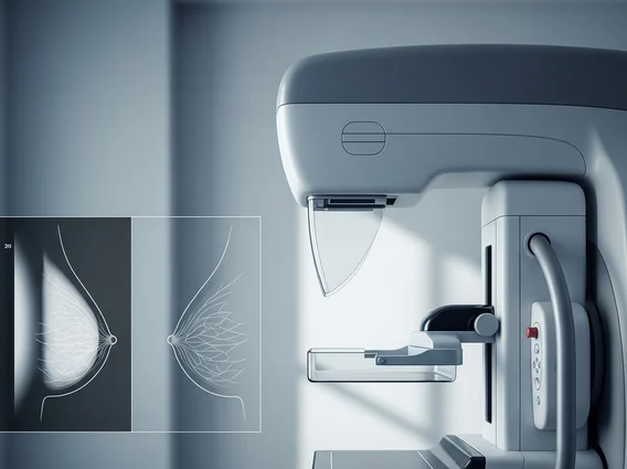
2 Dimensional Mammography (also known as 2D mammography) is a conventional X-ray imaging technique specifically designed for the detection of breast cancer and other breast abnormalities. It involves taking two standard views of each breast – typically a craniocaudal (top-to-bottom) view and a mediolateral oblique (side-angle) view. These images provide a flattened, two-dimensional representation of the breast tissue, allowing radiologists to identify potential areas of concern such as masses, calcifications, or architectural distortions. It has been the cornerstone of breast cancer screening programs for decades, significantly contributing to early detection and improved outcomes.
How 2D Mammography Works and Its Advantages
The process of 2D mammography involves a specialized X-ray machine that emits a low dose of radiation to create images of the breast. During the procedure, the breast is gently but firmly compressed between two plates. This compression is crucial as it helps to spread out the breast tissue, reduce the thickness of the breast, minimize motion blur, and decrease the amount of radiation needed. The X-ray beam then passes through the compressed breast, and the resulting image is captured on a detector. The entire process typically takes only a few minutes per breast.
The Benefits of 2D mammogram are numerous, making it a valuable tool in breast health. These advantages include:
- Widespread Availability: 2D mammography equipment is readily available in most healthcare facilities globally, making it accessible for routine screening.
- Cost-Effectiveness: It is generally less expensive than newer imaging modalities, which can be a significant factor for population-based screening programs.
- Established Efficacy: Decades of research and clinical use have demonstrated its effectiveness in detecting breast cancers, particularly larger or more obvious lesions.
- Lower Radiation Dose: While using X-rays, the radiation dose is kept as low as reasonably achievable, and the benefits of early detection far outweigh the minimal risks.
2D Mammography vs. 3D Mammography: Key Differences
The primary distinction between 2D mammography vs 3D mammography, also known as digital breast tomosynthesis (DBT), lies in how the images are acquired and presented. While 2D mammography captures a single, flattened image of the breast from two angles, 3D mammography takes multiple X-ray images from different angles around the breast. These images are then reconstructed by a computer to create a series of thin, cross-sectional slices of the breast tissue.
This multi-slice approach of 3D mammography significantly reduces the issue of tissue overlap, which is a common limitation in 2D mammography. In 2D images, dense breast tissue or overlapping normal structures can obscure abnormalities or create false alarms. 3D mammography allows radiologists to scroll through the breast tissue layer by layer, making it easier to distinguish between overlapping normal tissue and actual lesions. This often leads to improved cancer detection rates, especially in women with dense breasts, and can reduce the number of false positives, thereby decreasing recall rates for additional imaging. However, 3D mammography typically involves a slightly higher radiation dose and is not as universally available or covered by insurance as 2D mammography.