18f Naf Pet
18F NaF PET is a specialized medical imaging technique used primarily to assess bone metabolism and detect skeletal abnormalities. This advanced diagnostic tool offers detailed insights into bone activity, making it invaluable in various clinical settings.
Key Takeaways
- 18F NaF PET is a highly sensitive imaging technique for evaluating bone metabolism.
- It utilizes a radioactive tracer, sodium fluoride (18F-NaF), which accumulates in areas of increased bone turnover.
- The scan is particularly effective in detecting bone metastases, fractures, and other skeletal pathologies.
- It provides superior resolution and quantitative data compared to traditional bone scans.
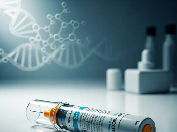
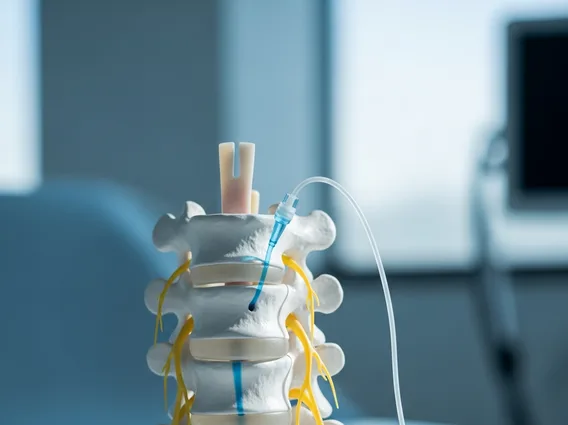
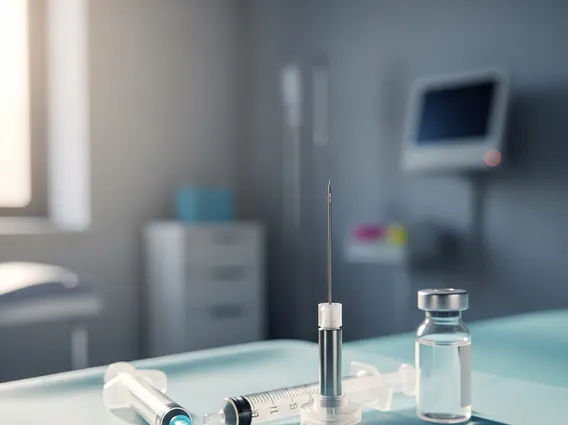
- The procedure involves intravenous injection of the tracer, followed by PET scanning to visualize tracer uptake.
What is 18f Naf Pet?
18F NaF PET refers to Positron Emission Tomography (PET) utilizing the radiotracer Fluorine-18 sodium fluoride (18F-NaF). This advanced nuclear medicine imaging technique is highly sensitive for detecting and characterizing changes in bone metabolism. Unlike traditional bone scans that primarily show blood flow and osteoblastic activity, an 18F NaF PET scan directly measures bone turnover, making it a powerful tool for identifying areas of increased bone formation or destruction. The radiotracer 18F-NaF behaves similarly to calcium, integrating into the bone matrix at sites of active bone remodeling. This allows for precise localization of skeletal abnormalities, often before they are visible on other imaging modalities.
How 18f Naf Pet Scans Work
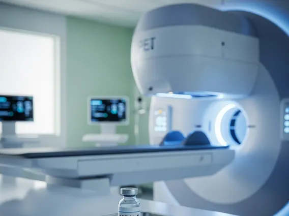
The mechanism of 18F NaF PET involves the intravenous administration of a small, safe dose of the radioactive tracer, 18F-NaF. Once injected, the tracer travels through the bloodstream and rapidly accumulates in areas of high bone metabolic activity, such as those associated with bone growth, repair, or disease. The fluoride ion (F-) exchanges with hydroxyl groups in the hydroxyapatite crystals of the bone, becoming incorporated into the bone matrix. After a waiting period, typically 60-90 minutes, to allow for tracer uptake into the bone and clearance from soft tissues, the patient undergoes a PET scan. The PET scanner detects the gamma rays emitted when positrons from the 18F isotope annihilate with electrons in the body. These signals are then processed by a computer to create detailed, three-dimensional images of the skeleton, highlighting areas of increased tracer uptake. This process is how 18F NaF PET imaging explained provides highly detailed and quantitative information about bone metabolism. The high spatial resolution of PET combined with the rapid uptake and clearance kinetics of 18F-NaF results in images with excellent contrast and diagnostic accuracy.
Clinical Uses of 18f Naf Pet Imaging
The clinical uses of 18F NaF PET imaging are diverse and primarily focused on evaluating skeletal health, particularly in oncology and orthopedics. Its high sensitivity makes it superior to conventional bone scintigraphy for detecting early bone metastases, which are a common complication of many cancers, including prostate, breast, and lung cancer. According to a study published in the Journal of Nuclear Medicine, 18F-NaF PET demonstrated higher sensitivity (96%) compared to bone scintigraphy (78%) for detecting bone metastases in prostate cancer patients. This early detection can significantly impact treatment planning and patient prognosis.
Other important clinical applications include:
- Detection of occult fractures: Identifying stress fractures or other subtle bone injuries that may not be apparent on X-rays or even MRI.
- Assessment of degenerative joint disease: Evaluating the extent of osteoarthritic changes and inflammatory processes in joints.
- Monitoring response to therapy: Tracking changes in bone lesions in response to cancer treatments or other interventions.
- Evaluation of metabolic bone diseases: Assisting in the diagnosis and management of conditions like Paget’s disease or osteomalacia.
- Pre-surgical planning: Providing detailed anatomical and metabolic information for complex orthopedic procedures.
The quantitative nature of 18F NaF PET allows clinicians to measure the rate of tracer uptake, offering objective data for assessing disease activity and treatment effectiveness.