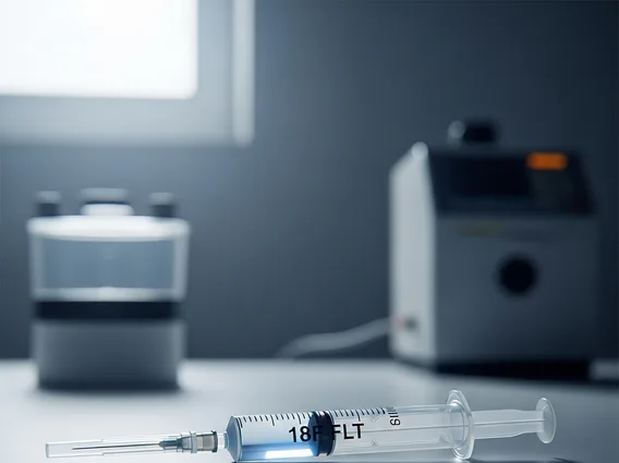
18f Flt
18F-FLT (Fluorothymidine) is a specialized radiotracer used in medical imaging, primarily with Positron Emission Tomography (PET) scans. It plays a crucial role in visualizing and assessing cellular proliferation, particularly in the context of oncology.
Key Takeaways
- 18F-FLT is a radiolabeled nucleoside analog used in PET imaging.
- It acts as a radiotracer information agent by indicating cellular proliferation.
- Primary 18F-FLT uses and applications are in oncology for tumor assessment.
- It helps monitor treatment response and distinguish tumor recurrence.
- 18F-FLT medical imaging provides insights into tumor biology beyond metabolic activity.
What is 18F-FLT (Fluorothymidine)?
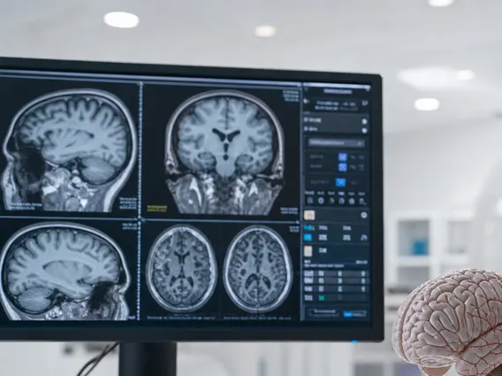
18F-FLT (Fluorothymidine) is a synthetic nucleoside analog labeled with the positron-emitting radioisotope fluorine-18. This compound is designed to be incorporated into the DNA synthesis pathway of cells, specifically reflecting the activity of thymidine kinase 1 (TK1), an enzyme upregulated during the S-phase of the cell cycle when DNA replication occurs. Unlike 18F-FDG, another widely used PET radiotracer that measures glucose metabolism (a general indicator of cellular activity), 18F-FLT directly assesses cellular proliferation. This distinction is crucial because some tumors may exhibit high metabolic activity without rapid proliferation, or vice versa. By specifically targeting the TK1 enzyme, 18F-FLT provides a more direct measure of the fraction of cells actively dividing, making it a valuable tool for understanding tumor growth dynamics and response to treatments that interfere with DNA synthesis.
As a radiotracer information agent, 18F-FLT is administered intravenously and then accumulates in cells undergoing active DNA synthesis. The fluorine-18 isotope emits positrons, which interact with electrons in the body, producing gamma rays detected by a PET scanner. This process allows clinicians to visualize and quantify areas of high cell proliferation, providing insights into the biological aggressiveness of tumors and their response to various therapies. Its mechanism offers a distinct advantage in situations where metabolic activity might not accurately reflect proliferative status, offering a more specific marker for tumor growth.
Applications of 18F-FLT in Medical Imaging
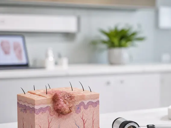
The primary 18F-FLT uses and applications are found within the field of oncology, where its unique ability to non-invasively measure cellular proliferation offers significant diagnostic, prognostic, and predictive value. 18F-FLT medical imaging is particularly useful for assessing tumor growth kinetics, understanding the biological aggressiveness of a malignancy, and monitoring the effectiveness of various cancer treatments, especially those designed to inhibit cell division or DNA replication.
Specific applications include:
- Monitoring Treatment Response: 18F-FLT PET scans can detect early changes in tumor proliferation following chemotherapy, radiation therapy, or targeted therapies. A significant decrease in 18F-FLT uptake can indicate a positive response to treatment, often before changes in tumor size are visible on conventional anatomical imaging, allowing for earlier adjustment of therapeutic strategies.
- Assessing Tumor Aggressiveness and Prognosis: Higher 18F-FLT uptake in a tumor often correlates with a more aggressive phenotype and faster growth rate. This information can aid clinicians in determining prognosis and guiding the intensity and type of treatment, contributing to personalized cancer management.
- Differentiating Tumor Recurrence from Post-Treatment Changes: In patients who have undergone treatment, it can be challenging to distinguish between residual or recurrent tumor and benign post-treatment changes like inflammation or necrosis. 18F-FLT can help in this differentiation, as recurrent tumors typically show increased proliferation, while benign changes generally do not.
- Guiding Biopsy and Treatment Planning: Areas of high 18F-FLT uptake can guide biopsy procedures to the most metabolically active and proliferative parts of a tumor, increasing the diagnostic yield. It can also inform radiation therapy planning by identifying regions of active tumor growth that require more focused treatment.
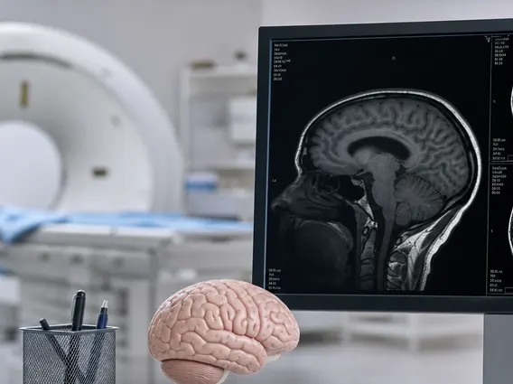
While predominantly used in oncology, ongoing research is exploring other potential applications where cell proliferation is a key pathological factor, such as in certain inflammatory conditions, infectious diseases, or even neurodegenerative disorders where glial cell activation is observed. However, its established clinical utility remains strongest and most validated in cancer management. It offers a crucial complementary perspective to other imaging modalities, such as 18F-FDG PET or anatomical MRI/CT scans, by providing direct insights into the fundamental process of cell division rather than just metabolic activity or structural changes. This makes 18F-FLT a powerful tool in the personalized medicine approach to cancer care.