
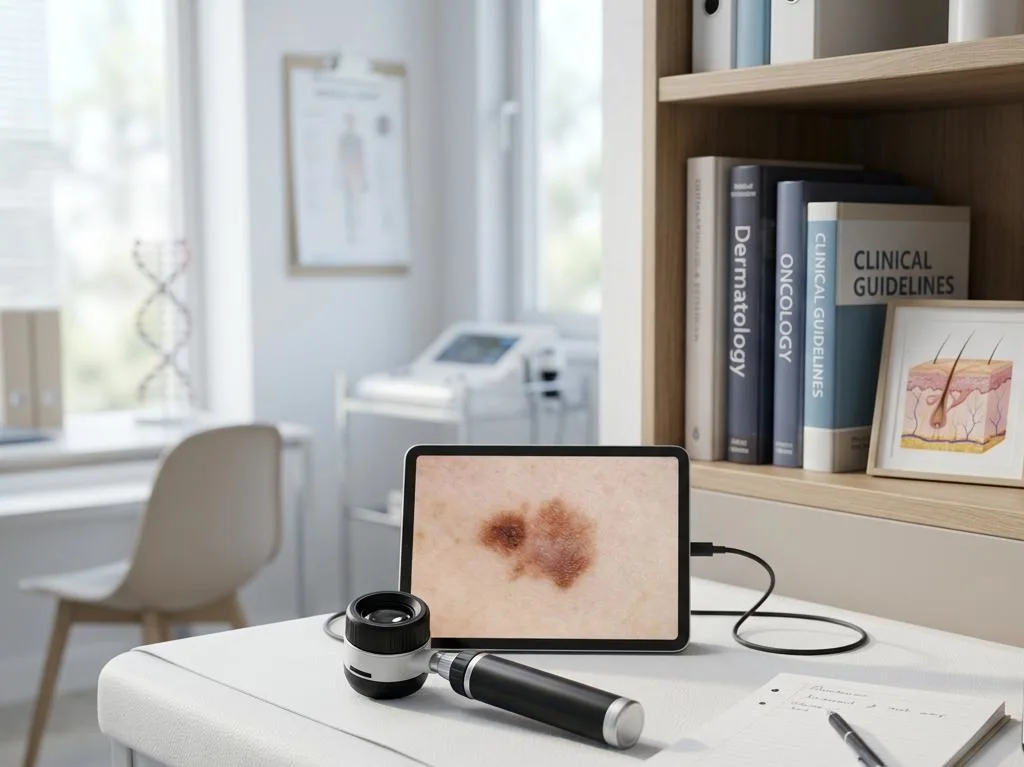
Risk Factors, Detection, Prognosis of Melanoma

Risk Factors, Detection, Prognosis of Melanoma

Risk Factors, Detection, Prognosis of Melanoma

Melanoma is one of the most serious forms of skin cancer, arising from the pigment-producing cells known as melanocytes. Understanding the risk factors, detection methods, and long-term outlook of this disease empowers patients and clinicians to act swiftly and make informed decisions.

Key Takeaways

  • UV radiation exposure and genetic predisposition are among the leading contributors to melanoma development.
  • The ABCDE criteria and regular skin examinations are foundational tools for early melanoma detection.
  • Melanoma is diagnosed through biopsy and staged using the TNM classification system.
  • Survival rates vary significantly by stage, with localized melanoma carrying a five-year survival rate exceeding 98%.
  • Early detection dramatically improves the long-term outlook for patients with melanoma.

Main Risk Factors for Developing Melanoma Skin Cancer

Melanoma risk factors and warning signs encompass a broad range of genetic, environmental, and behavioral elements that increase a person’s likelihood of developing this malignancy. Among the most significant is exposure to ultraviolet (UV) radiation, either from natural sunlight or artificial sources such as tanning beds. According to the World Health Organization (WHO), UV radiation is classified as a Group 1 carcinogen, meaning there is sufficient evidence that it causes cancer in humans. Individuals who experience repeated sunburns, particularly during childhood, face a substantially elevated risk.

Genetic factors also play a critical role. A family history of melanoma increases an individual’s risk two- to threefold compared to the general population. Mutations in specific genes, such as CDKN2A, are associated with hereditary melanoma syndromes. People with fair skin, light-colored eyes, and red or blonde hair have less melanin to protect against UV damage, making them inherently more vulnerable. Additionally, the presence of numerous or atypical moles — known as dysplastic nevi — is a well-established predictor of melanoma risk.

Immune suppression is another important consideration. Patients who have undergone organ transplantation or those living with conditions that compromise immune function face a higher risk of developing melanoma skin cancer. A personal history of any prior skin cancer further compounds this susceptibility. Age is also a contributing variable; although melanoma can occur at any age, its incidence increases significantly after age 50, and it remains one of the most common cancers diagnosed in adults under 30, according to the American Cancer Society.

  • Prolonged or intense UV radiation exposure (sun and tanning beds)
  • Fair skin, light eyes, or red/blonde hair
  • Family or personal history of melanoma or skin cancer
  • Large number of moles or presence of atypical (dysplastic) nevi
  • Weakened immune system due to disease or medication
  • Genetic mutations, particularly in CDKN2A

Early Detection Methods and Warning Signs of Melanoma

Identifying melanoma at its earliest stage is one of the most effective strategies for improving patient outcomes. The most widely used framework for recognizing suspicious lesions is the ABCDE criteria, which evaluates moles and skin spots based on Asymmetry, Border irregularity, Color variation, Diameter greater than six millimeters, and Evolution over time. Any lesion that changes in size, shape, or color warrants prompt evaluation by a dermatologist.

The early detection methods for melanoma available today extend well beyond visual inspection. Dermoscopy, a non-invasive technique that uses a handheld magnifying tool with polarized light, enables clinicians to examine subsurface skin structures not visible to the naked eye. Studies have shown that dermoscopy improves diagnostic accuracy by approximately 20–30% compared to unaided visual inspection. Total-body photography is another method used in high-risk individuals, providing a baseline record of the skin that allows clinicians to track changes over time.

Digital mole mapping, which combines total-body photography with dermatoscopic imaging, is increasingly used for surveillance in patients with multiple atypical nevi. Artificial intelligence–assisted image analysis has also emerged as a promising adjunct, with some algorithms demonstrating diagnostic accuracy comparable to experienced dermatologists. However, no technology replaces regular clinical skin examinations and patient self-monitoring. The American Academy of Dermatology recommends annual full-body skin checks for individuals at elevated risk, alongside monthly self-examinations at home.

Warning signs that should prompt immediate medical consultation include a new mole appearing after age 30, a lesion that bleeds or itches persistently, and any skin change that does not resolve within a few weeks. Patients with a known history of atypical moles or a family history of melanoma should adhere to a more rigorous surveillance schedule as directed by their dermatologist.

How Melanoma Is Diagnosed and Staged

A definitive melanoma diagnosis requires histopathological confirmation through a skin biopsy. When a suspicious lesion is identified, the preferred approach is an excisional biopsy, in which the entire lesion is removed with a small margin of surrounding tissue. This provides the pathologist with sufficient material to evaluate tumor thickness, ulceration, and mitotic rate — all of which are critical determinants of stage and prognosis.

Once a biopsy confirms malignancy, staging is performed using the TNM (Tumor, Node, Metastasis) classification system developed by the American Joint Committee on Cancer (AJCC). The T category reflects tumor thickness as measured by Breslow depth, along with the presence or absence of ulceration. The N category assesses regional lymph node involvement, while M indicates distant metastasis. Sentinel lymph node biopsy (SLNB) is commonly performed in patients with intermediate-thickness tumors to assess regional nodal spread without removing all surrounding lymph nodes.

Imaging studies such as positron emission tomography (PET) scans, computed tomography (CT), and magnetic resonance imaging (MRI) are employed when regional or distant spread is suspected. Molecular testing for mutations in genes such as BRAF, NRAS, and KIT is now a standard component of the diagnostic workup, as these findings directly guide targeted therapy decisions. The integration of genetic profiling into staging has markedly refined the precision of melanoma management over the past decade.

Melanoma Prognosis: Survival Rates and Long-Term Outlook by Stage

The melanoma prognosis by stage and survival rate varies considerably depending on how advanced the disease is at the time of diagnosis. Stage is the single most powerful predictor of outcome, and localized disease carries a far more favorable prognosis than metastatic melanoma. According to the National Cancer Institute’s Surveillance, Epidemiology, and End Results (SEER) database, the five-year relative survival rate for localized melanoma exceeds 98%, while regional disease drops to approximately 68%, and distant-stage melanoma falls to around 30%.

Beyond stage, several tumor characteristics influence the melanoma diagnosis and long-term outlook. Breslow depth — the vertical thickness of the tumor — is among the strongest prognostic indicators in early-stage disease. Thicker tumors are associated with a greater likelihood of lymph node involvement and distant metastasis. The presence of ulceration and an elevated mitotic rate are additional histologic features that independently worsen prognosis and are incorporated into AJCC staging criteria.

Stage Description 5-Year Survival Rate (Approximate)
Stage I Thin, localized tumor; no lymph node involvement 98–99%
Stage II Thicker tumor, possibly ulcerated; no nodal spread 80–90%
Stage III Regional lymph node involvement 60–70%
Stage IV Distant metastasis to organs or distant lymph nodes 15–30%

Advances in immunotherapy and targeted therapy have substantially improved outcomes for patients with advanced-stage melanoma. Immune checkpoint inhibitors such as pembrolizumab and nivolumab have demonstrated durable responses in a subset of Stage IV patients, with some achieving long-term remission. Targeted therapies addressing BRAF V600 mutations have similarly transformed the management of metastatic disease. Despite these improvements, long-term survival for Stage IV melanoma remains a challenge, reinforcing the irreplaceable value of early detection and prompt treatment.

Frequently Asked Questions

What distinguishes a normal mole from a potentially cancerous one?

Normal moles are typically symmetrical, have smooth and well-defined borders, display a uniform color, and remain stable over time. Concerning lesions often demonstrate asymmetry, irregular or notched borders, multiple shades of brown, black, red, or white, diameters larger than six millimeters, and noticeable changes in size or shape. Any mole that evolves, bleeds, or itches should be evaluated by a dermatologist promptly, as these changes may indicate early melanoma development.

Can melanoma develop in areas not exposed to the sun?

Yes. Although UV radiation is a primary driver, melanoma can arise in areas with little or no sun exposure, including the soles of the feet, palms, under the nails, and even the eyes or mucous membranes. These subtypes, such as acral lentiginous melanoma, are more common in individuals with darker skin tones and are often diagnosed at a later stage due to their atypical locations, underscoring the need for full-body skin surveillance regardless of skin type.

Does a family history of melanoma require special monitoring?

A family history of melanoma significantly elevates personal risk and typically warrants a more structured surveillance program. Individuals with one or more first-degree relatives diagnosed with melanoma are advised to begin regular dermatological check-ups early in adulthood. Genetic counseling may also be recommended to assess for hereditary mutations. Clinicians often use total-body photography or digital mole mapping in these patients to detect subtle changes that may not be apparent during routine examination.


[EN] Cancer Types
Cancer Clinical Trial Options

Specialized matching specifically for oncology clinical trials and cancer care research.

Your Birthday


By filling out this form, you're consenting only to release your medical records. You're not agreeing to participate in clinical trials yet.

Melanoma is one of the most serious forms of skin cancer, arising from the pigment-producing cells known as melanocytes. Understanding the risk factors, detection methods, and long-term outlook of this disease empowers patients and clinicians to act swiftly and make informed decisions.

Key Takeaways

  • UV radiation exposure and genetic predisposition are among the leading contributors to melanoma development.
  • The ABCDE criteria and regular skin examinations are foundational tools for early melanoma detection.
  • Melanoma is diagnosed through biopsy and staged using the TNM classification system.
  • Survival rates vary significantly by stage, with localized melanoma carrying a five-year survival rate exceeding 98%.
  • Early detection dramatically improves the long-term outlook for patients with melanoma.

Main Risk Factors for Developing Melanoma Skin Cancer

Melanoma risk factors and warning signs encompass a broad range of genetic, environmental, and behavioral elements that increase a person’s likelihood of developing this malignancy. Among the most significant is exposure to ultraviolet (UV) radiation, either from natural sunlight or artificial sources such as tanning beds. According to the World Health Organization (WHO), UV radiation is classified as a Group 1 carcinogen, meaning there is sufficient evidence that it causes cancer in humans. Individuals who experience repeated sunburns, particularly during childhood, face a substantially elevated risk.

Genetic factors also play a critical role. A family history of melanoma increases an individual’s risk two- to threefold compared to the general population. Mutations in specific genes, such as CDKN2A, are associated with hereditary melanoma syndromes. People with fair skin, light-colored eyes, and red or blonde hair have less melanin to protect against UV damage, making them inherently more vulnerable. Additionally, the presence of numerous or atypical moles — known as dysplastic nevi — is a well-established predictor of melanoma risk.

Immune suppression is another important consideration. Patients who have undergone organ transplantation or those living with conditions that compromise immune function face a higher risk of developing melanoma skin cancer. A personal history of any prior skin cancer further compounds this susceptibility. Age is also a contributing variable; although melanoma can occur at any age, its incidence increases significantly after age 50, and it remains one of the most common cancers diagnosed in adults under 30, according to the American Cancer Society.

  • Prolonged or intense UV radiation exposure (sun and tanning beds)
  • Fair skin, light eyes, or red/blonde hair
  • Family or personal history of melanoma or skin cancer
  • Large number of moles or presence of atypical (dysplastic) nevi
  • Weakened immune system due to disease or medication
  • Genetic mutations, particularly in CDKN2A

Early Detection Methods and Warning Signs of Melanoma

Identifying melanoma at its earliest stage is one of the most effective strategies for improving patient outcomes. The most widely used framework for recognizing suspicious lesions is the ABCDE criteria, which evaluates moles and skin spots based on Asymmetry, Border irregularity, Color variation, Diameter greater than six millimeters, and Evolution over time. Any lesion that changes in size, shape, or color warrants prompt evaluation by a dermatologist.

The early detection methods for melanoma available today extend well beyond visual inspection. Dermoscopy, a non-invasive technique that uses a handheld magnifying tool with polarized light, enables clinicians to examine subsurface skin structures not visible to the naked eye. Studies have shown that dermoscopy improves diagnostic accuracy by approximately 20–30% compared to unaided visual inspection. Total-body photography is another method used in high-risk individuals, providing a baseline record of the skin that allows clinicians to track changes over time.

Digital mole mapping, which combines total-body photography with dermatoscopic imaging, is increasingly used for surveillance in patients with multiple atypical nevi. Artificial intelligence–assisted image analysis has also emerged as a promising adjunct, with some algorithms demonstrating diagnostic accuracy comparable to experienced dermatologists. However, no technology replaces regular clinical skin examinations and patient self-monitoring. The American Academy of Dermatology recommends annual full-body skin checks for individuals at elevated risk, alongside monthly self-examinations at home.

Warning signs that should prompt immediate medical consultation include a new mole appearing after age 30, a lesion that bleeds or itches persistently, and any skin change that does not resolve within a few weeks. Patients with a known history of atypical moles or a family history of melanoma should adhere to a more rigorous surveillance schedule as directed by their dermatologist.

How Melanoma Is Diagnosed and Staged

A definitive melanoma diagnosis requires histopathological confirmation through a skin biopsy. When a suspicious lesion is identified, the preferred approach is an excisional biopsy, in which the entire lesion is removed with a small margin of surrounding tissue. This provides the pathologist with sufficient material to evaluate tumor thickness, ulceration, and mitotic rate — all of which are critical determinants of stage and prognosis.

Once a biopsy confirms malignancy, staging is performed using the TNM (Tumor, Node, Metastasis) classification system developed by the American Joint Committee on Cancer (AJCC). The T category reflects tumor thickness as measured by Breslow depth, along with the presence or absence of ulceration. The N category assesses regional lymph node involvement, while M indicates distant metastasis. Sentinel lymph node biopsy (SLNB) is commonly performed in patients with intermediate-thickness tumors to assess regional nodal spread without removing all surrounding lymph nodes.

Imaging studies such as positron emission tomography (PET) scans, computed tomography (CT), and magnetic resonance imaging (MRI) are employed when regional or distant spread is suspected. Molecular testing for mutations in genes such as BRAF, NRAS, and KIT is now a standard component of the diagnostic workup, as these findings directly guide targeted therapy decisions. The integration of genetic profiling into staging has markedly refined the precision of melanoma management over the past decade.

Melanoma Prognosis: Survival Rates and Long-Term Outlook by Stage

The melanoma prognosis by stage and survival rate varies considerably depending on how advanced the disease is at the time of diagnosis. Stage is the single most powerful predictor of outcome, and localized disease carries a far more favorable prognosis than metastatic melanoma. According to the National Cancer Institute’s Surveillance, Epidemiology, and End Results (SEER) database, the five-year relative survival rate for localized melanoma exceeds 98%, while regional disease drops to approximately 68%, and distant-stage melanoma falls to around 30%.

Beyond stage, several tumor characteristics influence the melanoma diagnosis and long-term outlook. Breslow depth — the vertical thickness of the tumor — is among the strongest prognostic indicators in early-stage disease. Thicker tumors are associated with a greater likelihood of lymph node involvement and distant metastasis. The presence of ulceration and an elevated mitotic rate are additional histologic features that independently worsen prognosis and are incorporated into AJCC staging criteria.

Stage Description 5-Year Survival Rate (Approximate)
Stage I Thin, localized tumor; no lymph node involvement 98–99%
Stage II Thicker tumor, possibly ulcerated; no nodal spread 80–90%
Stage III Regional lymph node involvement 60–70%
Stage IV Distant metastasis to organs or distant lymph nodes 15–30%

Advances in immunotherapy and targeted therapy have substantially improved outcomes for patients with advanced-stage melanoma. Immune checkpoint inhibitors such as pembrolizumab and nivolumab have demonstrated durable responses in a subset of Stage IV patients, with some achieving long-term remission. Targeted therapies addressing BRAF V600 mutations have similarly transformed the management of metastatic disease. Despite these improvements, long-term survival for Stage IV melanoma remains a challenge, reinforcing the irreplaceable value of early detection and prompt treatment.

Frequently Asked Questions

What distinguishes a normal mole from a potentially cancerous one?

Normal moles are typically symmetrical, have smooth and well-defined borders, display a uniform color, and remain stable over time. Concerning lesions often demonstrate asymmetry, irregular or notched borders, multiple shades of brown, black, red, or white, diameters larger than six millimeters, and noticeable changes in size or shape. Any mole that evolves, bleeds, or itches should be evaluated by a dermatologist promptly, as these changes may indicate early melanoma development.

Can melanoma develop in areas not exposed to the sun?

Yes. Although UV radiation is a primary driver, melanoma can arise in areas with little or no sun exposure, including the soles of the feet, palms, under the nails, and even the eyes or mucous membranes. These subtypes, such as acral lentiginous melanoma, are more common in individuals with darker skin tones and are often diagnosed at a later stage due to their atypical locations, underscoring the need for full-body skin surveillance regardless of skin type.

Does a family history of melanoma require special monitoring?

A family history of melanoma significantly elevates personal risk and typically warrants a more structured surveillance program. Individuals with one or more first-degree relatives diagnosed with melanoma are advised to begin regular dermatological check-ups early in adulthood. Genetic counseling may also be recommended to assess for hereditary mutations. Clinicians often use total-body photography or digital mole mapping in these patients to detect subtle changes that may not be apparent during routine examination.

[EN] Cancer Types
Cancer Clinical Trial Options

Specialized matching specifically for oncology clinical trials and cancer care research.

Your Birthday


By filling out this form, you're consenting only to release your medical records. You're not agreeing to participate in clinical trials yet.

Massive Bio has onboarded over 160,000+ cancer patients to find their clinical trial

Most Recent Article
















