
Radionuclide Scanning
Radionuclide scanning is a sophisticated medical imaging technique that provides valuable insights into the function and structure of organs and tissues within the body. It plays a crucial role in diagnosing and monitoring a wide range of medical conditions.
Key Takeaways
- Radionuclide scanning uses small, safe amounts of radioactive materials (radiotracers) to visualize organ function.
- These scans detect gamma rays emitted by radiotracers, which are then converted into detailed images by specialized cameras.
- The technique is non-invasive and provides unique functional information that other imaging methods may not capture.
- It is widely used across various medical specialties for diagnosing diseases affecting bones, heart, thyroid, and more.
- Different radiotracers and imaging protocols are employed depending on the specific organ or condition being investigated.
What is Radionuclide Scanning?
Radionuclide Scanning is a specialized branch of medical imaging, often referred to as nuclear medicine, that uses small, safe amounts of radioactive materials, known as radiotracers, to diagnose and assess various diseases. Unlike X-rays or CT scans, which primarily show anatomical structures, radionuclide scanning provides functional information by showing how organs and tissues are working at a molecular level. This allows clinicians to detect diseases in their early stages, sometimes even before structural changes are visible on other imaging tests. The radiotracers are designed to accumulate in specific organs or tissues, enabling targeted imaging.
This diagnostic approach is non-invasive and typically involves injecting a radiotracer into a vein, which then travels through the bloodstream to the target area. The amount of radiation used is very small and generally considered safe for diagnostic purposes, comparable to that received from other common imaging procedures. According to major health organizations, nuclear medicine procedures are essential tools in modern healthcare, contributing significantly to patient care by guiding treatment decisions and monitoring disease progression effectively.
How Radionuclide Scans Work
The fundamental principle behind how does radionuclide scan work involves the administration of a radiotracer and the subsequent detection of emitted radiation. Once introduced into the body, the radiotracer travels to the specific organ or tissue being examined, where it is absorbed or metabolized. The choice of radiotracer depends on the organ or process being studied; for instance, different tracers are used for bone scans versus cardiac scans.
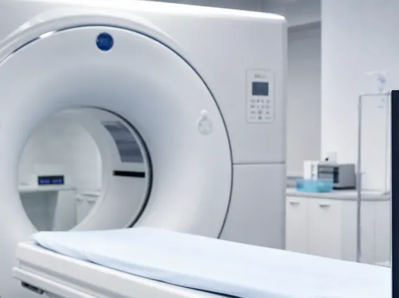
As the radiotracer decays, it emits gamma rays. These gamma rays are detected by a specialized external camera, known as a gamma camera or SPECT (Single-Photon Emission Computed Tomography) scanner, or a PET (Positron Emission Tomography) scanner. The camera does not emit radiation but rather captures the emitted signals. A computer then processes these signals to create detailed 2D or 3D images that illustrate the distribution and concentration of the radiotracer within the body. Areas with higher uptake of the radiotracer may indicate increased metabolic activity, inflammation, or the presence of tumors, while areas with lower uptake might suggest reduced function or tissue damage.
The process generally follows these steps:
- Radiotracer Administration: A small dose of a specific radiotracer is typically injected intravenously, though it can also be inhaled or swallowed depending on the scan type.
- Uptake and Distribution: The radiotracer travels through the bloodstream and accumulates in the target organ or tissue.
- Emission Detection: As the radiotracer decays, it emits gamma rays, which pass out of the body.
- Image Capture: A gamma camera or PET scanner detects these gamma rays from various angles.
- Image Reconstruction: A computer processes the detected signals to generate detailed images that show the functional activity of the scanned area.
Uses and Types of Radionuclide Imaging
Radionuclide scanning uses are extensive, providing crucial diagnostic information across numerous medical specialties. This imaging technique is invaluable for assessing organ function, detecting tumors, identifying infections, and evaluating blood flow. It offers a unique perspective on physiological processes that often cannot be obtained through other imaging modalities.
There are several types of radionuclide imaging, each tailored to specific diagnostic needs. These range from assessing bone health to evaluating cardiac function and identifying thyroid disorders. The versatility of radionuclide scanning makes it a cornerstone in the diagnosis and management of many complex conditions. For example, bone scans can detect fractures, infections, and cancers that might not be visible on conventional X-rays, while cardiac scans can assess blood flow to the heart muscle and identify areas of damage.
| Type of Scan | Primary Use | Radiotracer Example |
|---|---|---|
| Bone Scan | Detecting fractures, infections, arthritis, and bone cancer metastases. | Technetium-99m MDP |
| Cardiac Scan (Myocardial Perfusion Imaging) | Assessing blood flow to the heart muscle, identifying coronary artery disease and heart damage. | Technetium-99m Sestamibi or Thallium-201 |
| Thyroid Scan | Evaluating thyroid gland function, detecting nodules, hyperthyroidism, or hypothyroidism. | Iodine-123 or Technetium-99m Pertechnetate |
| Kidney Scan (Renal Scintigraphy) | Assessing kidney function, blood flow, and identifying obstructions or abnormalities. | Technetium-99m MAG3 or DTPA |
| PET Scan (Positron Emission Tomography) | Detecting cancer, assessing treatment response, evaluating brain disorders (e.g., Alzheimer’s), and heart conditions. | Fluorodeoxyglucose (FDG) labeled with Fluorine-18 |
These diverse applications highlight the critical role of radionuclide scanning in providing functional insights that complement anatomical imaging, leading to more accurate diagnoses and personalized treatment plans for patients.



















