
Radioactive Glucose
Radioactive glucose is a specialized medical tracer utilized in advanced imaging techniques to visualize metabolic activity within the body. This diagnostic tool plays a crucial role in the detection and management of various diseases, particularly in oncology, neurology, and cardiology.
Key Takeaways
- Radioactive glucose is a modified sugar molecule containing a radioactive isotope, primarily used in Positron Emission Tomography (PET) scans.
- It acts as a tracer, highlighting areas of high metabolic activity, which is characteristic of rapidly growing cells like those found in tumors.
- Medical applications include detecting and staging cancers, assessing treatment effectiveness, and diagnosing certain neurological and cardiac conditions.
- While it involves radiation exposure, the dose is minimal and considered safe for diagnostic purposes, with benefits typically outweighing the risks.
- Patients undergo specific preparation, such as fasting, to ensure accurate imaging results.
What is Radioactive Glucose?
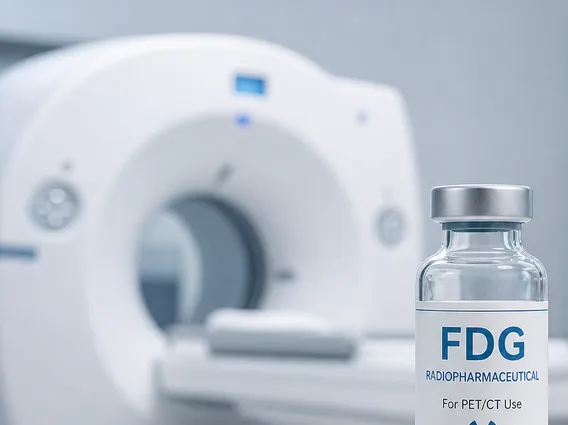
Radioactive Glucose refers to a glucose molecule that has been tagged with a small amount of a radioactive isotope, most commonly fluorine-18 (F-18). This modified sugar, often known as F-18 fluorodeoxyglucose (FDG), is administered to patients as a radiopharmaceutical. Its primary purpose is to act as a tracer in medical imaging, specifically for Positron Emission Tomography (PET) scans. The fundamental principle behind its use is that cells in the body, particularly those that are rapidly growing or highly active, consume glucose at an accelerated rate. By introducing a radioactive form of glucose, medical professionals can observe and map these areas of increased metabolic demand.
Once injected into the bloodstream, radioactive glucose travels throughout the body and is absorbed by cells in a manner similar to regular glucose. However, unlike normal glucose, the radioactive glucose molecule cannot be fully metabolized past a certain point, causing it to accumulate within metabolically active cells. This accumulation allows the radioactive isotope to emit positrons, which are then detected by the PET scanner, creating detailed images of tissue function and metabolic activity.
How Radioactive Glucose Works and Its Medical Applications
When administered, radioactive glucose is absorbed by cells throughout the body, with a higher concentration accumulating in areas of increased metabolic activity. The F-18 isotope within the glucose molecule emits positrons, which quickly collide with electrons in the surrounding tissue, leading to the emission of gamma rays. These gamma rays are detected by the PET scanner, which then reconstructs a three-dimensional image showing where the radioactive glucose has accumulated. This process allows clinicians to visualize and quantify glucose metabolism in various tissues and organs, providing insights into their functional status.
The primary radioactive glucose medical applications are diverse and critical for modern diagnostics. It is extensively used in:
- Oncology: Identifying cancerous tumors, staging cancer, determining if cancer has spread (metastasis), monitoring the effectiveness of cancer treatments, and detecting cancer recurrence. Cancer cells typically have a higher metabolic rate and thus absorb more radioactive glucose than healthy cells.
- Neurology: Diagnosing conditions such as Alzheimer’s disease, epilepsy, and other brain disorders. PET scans with radioactive glucose can reveal areas of reduced glucose metabolism in the brain, which are indicative of neuronal dysfunction or degeneration.
- Cardiology: Assessing myocardial viability after a heart attack. It helps distinguish between damaged heart muscle that is still alive but not functioning well (hibernating myocardium) and irreversibly scarred tissue, guiding decisions about revascularization procedures.
This imaging technique provides functional information that complements anatomical imaging methods like CT or MRI, offering a more complete picture of disease processes.
Dangers and Safety of Radioactive Glucose
While radioactive glucose is a powerful diagnostic tool, concerns about the dangers of radioactive glucose primarily revolve around radiation exposure. However, the amount of radiation involved in a typical PET scan is carefully controlled and generally considered low for diagnostic purposes. The radioactive isotope, F-18, has a short half-life of approximately 110 minutes, meaning it decays rapidly and is quickly eliminated from the body, minimizing the patient’s overall radiation dose.
Safety protocols are rigorously followed to ensure patient well-being. Patients are typically advised to fast for several hours before the scan to optimize glucose uptake by target cells and to minimize background glucose levels. After the scan, patients are often encouraged to drink plenty of fluids to help flush the remaining radioactive tracer from their system. The effective radiation dose from a typical PET scan using radioactive glucose (F-18 FDG) is generally low, often ranging from 2 to 7 mSv, which is comparable to the annual background radiation exposure in some regions or other common diagnostic imaging procedures like a CT scan of the abdomen and pelvis. (Source: Radiological Society of North America (RSNA)).
For the vast majority of patients, the diagnostic benefits of using radioactive glucose in PET imaging significantly outweigh the minimal risks associated with radiation exposure. The information gained from these scans can be critical for accurate diagnosis, staging, and treatment planning, ultimately leading to improved patient outcomes.



















