Papillary Reticular Dermal Interface
The papillary reticular dermal interface represents a critical anatomical and functional boundary within the skin, essential for maintaining its integrity and diverse physiological roles. This intricate zone facilitates vital interactions between the epidermis and dermis, impacting skin health and appearance.
Key Takeaways
- The papillary reticular dermal interface definition refers to the complex boundary between the superficial papillary dermis and the deeper reticular dermis.
- This interface is crucial for nutrient exchange, waste removal, and mechanical stability of the skin.
- It consists of two distinct layers: the papillary dermis, characterized by dermal papillae, and the reticular dermis, known for its dense collagen network.
- The structural integrity of this interface is vital for skin elasticity, strength, and overall function.
- Disruptions to this interface can lead to various dermatological conditions and impact wound healing.
What is the Papillary Reticular Dermal Interface?
The papillary reticular dermal interface is the transitional zone that separates the more superficial papillary layer of the dermis from its deeper, denser reticular layer. This interface is not merely a line but a complex region characterized by a gradual change in tissue composition and organization. It plays a fundamental role in anchoring the epidermis to the dermis, providing structural support, and enabling efficient communication between these two primary skin layers. Understanding this interface is crucial for comprehending skin physiology, pathology, and the mechanisms of aging and disease.
This critical boundary ensures the skin’s resilience and functional capacity. Its intricate architecture allows for the dynamic processes necessary for skin health, including cellular nourishment, waste product removal, and the transmission of sensory information. The integrity of this interface is paramount for maintaining the skin’s barrier function and its ability to withstand mechanical stress.
Structure of the Papillary Reticular Dermis
The structure of papillary reticular dermis is characterized by two distinct yet interconnected layers, each contributing uniquely to the overall function of the skin. These layers differ in their cellularity, fiber density, and vascularity, creating a gradient that supports various skin functions.
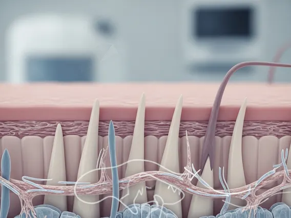
- Papillary Dermis: This is the superficial layer, directly beneath the epidermis, characterized by numerous finger-like projections called dermal papillae. These papillae interdigitate with epidermal ridges, significantly increasing the surface area for nutrient and waste exchange. The papillary dermis consists of loose connective tissue, containing fine collagen and elastic fibers, fibroblasts, mast cells, and a rich capillary network. It is also abundant in sensory nerve endings, contributing to touch and pain perception.
- Reticular Dermis: Lying deeper than the papillary dermis, the reticular dermis is composed of dense irregular connective tissue. It features thick bundles of collagen fibers (primarily type I) arranged in a coarse, interwoven network, along with robust elastic fibers. This layer provides the skin with its primary tensile strength and elasticity. It contains fewer cells than the papillary dermis but houses larger blood vessels, nerves, hair follicles, sebaceous glands, and sweat glands.
The interface between these two layers is a gradual transition zone where the fine fibers of the papillary dermis begin to merge with the thicker, more organized bundles of the reticular dermis. This structural continuum is essential for distributing mechanical forces across the dermis and maintaining overall skin integrity.
Function of the Dermal Interface
The papillary reticular dermal interface function is multifaceted, serving several vital roles that are indispensable for skin health and overall bodily well-being. Its primary functions revolve around mechanical support, nutrient exchange, and sensory perception.
Firstly, it provides robust mechanical support, anchoring the epidermis firmly to the underlying dermis. The interdigitating nature of the dermal papillae and epidermal ridges at this interface significantly increases the resistance to shearing forces, preventing epidermal detachment. This mechanical stability is crucial for the skin’s role as a protective barrier against physical trauma.
Secondly, this interface is a hub for nutrient and waste exchange. The rich capillary network within the papillary dermis facilitates the diffusion of oxygen and nutrients from the blood to the avascular epidermis, while simultaneously removing metabolic waste products. This efficient transport system is fundamental for epidermal cell proliferation, differentiation, and overall metabolic activity.
Furthermore, the dermal interface contributes significantly to sensory perception. The papillary dermis contains numerous Meissner’s corpuscles and free nerve endings, which are responsible for detecting light touch, pressure, and temperature changes. This allows the skin to act as a crucial sensory organ, relaying information about the external environment to the nervous system. The structural and functional integrity of this interface is therefore critical for maintaining healthy skin and its diverse physiological roles.