Magnetic Resonance Perfusion Imaging
Magnetic Resonance Perfusion Imaging (MRPI) is an advanced diagnostic technique that provides crucial insights into tissue blood flow and microvascular integrity. This non-invasive method is vital for assessing various medical conditions by visualizing the delivery of blood to tissues at a cellular level.
Key Takeaways
- Magnetic Resonance Perfusion Imaging (MRPI) measures blood flow to tissues, offering detailed hemodynamic information.
- It works by tracking the passage of a contrast agent through the bloodstream, revealing how well tissues are perfused.
- MRPI is widely used in neurology and oncology for diagnosing and monitoring conditions like stroke, tumors, and neurodegenerative diseases.
- The technique provides non-invasive, high-resolution images, aiding in early detection, treatment planning, and evaluating therapy response.
- Its ability to differentiate between viable and damaged tissue makes it an invaluable tool in clinical decision-making.
What is Magnetic Resonance Perfusion Imaging (MRPI)?
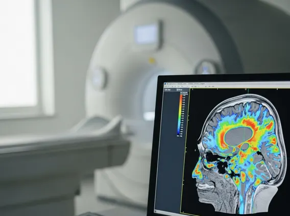
Magnetic Resonance Perfusion Imaging (MRPI) is a specialized magnetic resonance imaging technique that measures the amount of blood flowing through the capillaries in a specific tissue over time. This imaging modality provides functional information about tissue perfusion, which is the process of blood delivering oxygen and nutrients to the capillary beds of biological tissue. By quantifying parameters such as cerebral blood flow (CBF), cerebral blood volume (CBV), and mean transit time (MTT), MRPI helps clinicians understand the physiological state of tissues, particularly in the brain and other organs. It is essential for identifying areas of compromised blood supply, which can indicate various pathological conditions.
The technique relies on the rapid acquisition of MR images during the passage of a contrast agent, typically a gadolinium-based compound, through the vascular system. This allows for dynamic assessment of tissue microcirculation, making it a powerful tool for evaluating conditions where blood supply is critical.
How Magnetic Resonance Perfusion Imaging Works
Magnetic Resonance Perfusion Imaging operates by detecting changes in the magnetic signal as a contrast agent passes through the body’s tissues. When a gadolinium-based contrast agent is injected intravenously, it travels through the bloodstream, altering the local magnetic field. MR scanners are designed to capture these transient signal changes with high temporal resolution. As the contrast agent enters and exits the capillaries of a tissue, it causes a temporary decrease in the MR signal intensity, which is then measured and analyzed.
The data collected during this dynamic process is used to generate quantitative maps of perfusion parameters. These maps illustrate the distribution and kinetics of blood flow, allowing radiologists to visualize areas of reduced or increased perfusion. For instance, in the brain, areas with restricted blood flow due to a stroke will show different perfusion characteristics compared to healthy tissue. The precise mechanism involves the T2* shortening effect of the paramagnetic contrast agent, which is sensitive to its concentration within the microvasculature. Advanced computational models then process these signal changes to derive clinically relevant perfusion metrics.
Applications and Benefits of MR Perfusion Imaging
Magnetic Resonance Perfusion Imaging applications are diverse, primarily concentrated in neurology and oncology, but also extending to cardiac and renal imaging. In neuroimaging, MRPI is indispensable for the acute assessment of stroke, helping to distinguish between salvageable brain tissue (penumbra) and irreversibly damaged tissue (infarct core). This distinction is crucial for guiding thrombolytic therapy decisions. It is also used in the evaluation of brain tumors, aiding in grading, differentiating tumor recurrence from radiation necrosis, and monitoring response to treatment by assessing tumor angiogenesis.
The benefits of MR perfusion imaging are substantial, offering a non-invasive method to obtain functional information that complements structural imaging. Key advantages include:
- Early Detection: Can identify subtle changes in blood flow indicative of disease processes before structural changes are apparent.
- Treatment Guidance: Provides critical information for planning interventions, such as determining the optimal timing for stroke treatment or guiding biopsy locations in tumors.
- Prognostic Value: Perfusion parameters can offer insights into disease progression and patient outcomes.
- Monitoring Therapy Response: Allows for the objective assessment of how well a treatment is working by observing changes in tissue perfusion.
- Reduced Radiation Exposure: Unlike some other perfusion imaging techniques (e.g., CT perfusion), MRPI does not involve ionizing radiation, making it safer for repeated studies.
MRPI’s ability to provide detailed, quantitative information about tissue microcirculation makes it an invaluable tool for diagnosis, treatment planning, and patient management across various medical specialties.