
Karyotyping
Karyotyping is a laboratory technique used to examine an individual’s set of chromosomes. This genetic test helps identify chromosomal abnormalities that can be associated with various genetic disorders, developmental delays, or reproductive issues.
Key Takeaways
- Karyotyping is a genetic test that analyzes the number and structure of an individual’s chromosomes.
- It is used to diagnose chromosomal disorders, investigate causes of developmental delays, and assess recurrent miscarriages.
- The procedure involves culturing cells, arresting them during division, and then staining and photographing the chromosomes to create a karyogram.
- Results are interpreted by comparing the patient’s karyogram to a normal human karyotype, identifying numerical or structural anomalies.
- Common findings include aneuploidies like Down syndrome, or structural changes such as translocations and deletions.
What Is Karyotyping?
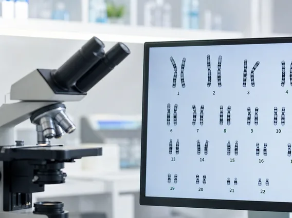
Karyotyping is a laboratory technique that provides a visual representation of an individual’s chromosomes. It involves arranging a complete set of chromosomes from a cell, typically during metaphase of cell division, into a standardized format called a karyogram. This process allows geneticists to examine the number, size, shape, and banding patterns of chromosomes to detect any abnormalities. Each human cell normally contains 23 pairs of chromosomes, totaling 46, with 22 pairs of autosomes and one pair of sex chromosomes (XX for females, XY for males).
Purpose and Procedure of Karyotyping
The purpose of karyotyping test is primarily to diagnose chromosomal disorders that can lead to various health conditions. This test is often recommended for individuals with unexplained developmental delays, intellectual disabilities, congenital anomalies, or for couples experiencing recurrent miscarriages or infertility. For instance, approximately 1 in 160 live births involves a chromosomal abnormality, many of which can be detected by karyotyping (Source: National Institutes of Health).
The karyotyping procedure explained involves several key steps:
- Sample Collection: A sample of cells is collected, most commonly from blood (lymphocytes), but also from bone marrow, amniotic fluid (amniocentesis), or chorionic villus tissue (CVS).
- Cell Culture: The collected cells are cultured in a laboratory to stimulate growth and division.
- Cell Arrest: Cells are treated with a chemical (e.g., colchicine) to arrest them in metaphase, the stage of cell division where chromosomes are most condensed and visible.
- Staining and Imaging: The cells are then spread onto a slide, stained (often with Giemsa stain to produce characteristic banding patterns), and viewed under a microscope. High-resolution images are captured.
- Karyogram Creation: The chromosomes from the images are cut out (digitally or manually) and arranged in homologous pairs, ordered by size, from largest to smallest (1 to 22), followed by the sex chromosomes.
Interpreting Karyotyping Results
The karyotyping results interpretation involves a careful analysis of the created karyogram. Geneticists examine the total number of chromosomes and look for any structural changes within them. A normal human karyotype is typically reported as 46,XX for females and 46,XY for males. Any deviation from this standard indicates a chromosomal abnormality.
Common types of abnormalities identified through karyotyping include:
- Numerical Abnormalities (Aneuploidy): These involve an extra or missing chromosome.
- Trisomy: Presence of an extra copy of a chromosome (e.g., Trisomy 21 for Down syndrome, 47,XX,+21 or 47,XY,+21). Down syndrome affects about 1 in 700 babies born in the United States each year (Source: CDC).
- Monosomy: Absence of one chromosome from a pair (e.g., Monosomy X for Turner syndrome, 45,X).
- Structural Abnormalities: These involve changes in the structure of one or more chromosomes.
- Deletions: A portion of a chromosome is missing.
- Duplications: A portion of a chromosome is duplicated, resulting in extra genetic material.
- Translocations: A segment of one chromosome breaks off and attaches to another chromosome. These can be balanced (no net loss or gain of genetic material) or unbalanced.
- Inversions: A segment of a chromosome is reversed end-to-end.
- Rings: A chromosome breaks in two places and its ends fuse to form a ring.
Understanding these results is crucial for genetic counseling, allowing individuals and families to make informed decisions regarding medical management, family planning, and prognosis.



















