High Resolution Micro Ultrasound
High Resolution Micro Ultrasound (HRMUS) represents a significant advancement in medical imaging, offering unprecedented detail for visualizing small anatomical structures and subtle tissue changes. This innovative technology provides clinicians with a powerful tool for enhanced diagnostic accuracy across various medical specialties.
Key Takeaways
- High Resolution Micro Ultrasound (HRMUS) is an advanced imaging technique utilizing higher frequencies for superior detail.
- It works by employing high-frequency transducers (typically 20-70 MHz) to generate shorter sound waves, resulting in finer spatial resolution.
- The primary benefits of high resolution ultrasound imaging include improved diagnostic accuracy and earlier detection of subtle abnormalities.
- Key applications of micro ultrasound technology span areas like prostate imaging, dermatology, and musculoskeletal assessment.
- HRMUS is non-invasive and provides real-time imaging without ionizing radiation.
What is High Resolution Micro Ultrasound (HRMUS)?
High Resolution Micro Ultrasound (HRMUS) is an advanced medical imaging modality that utilizes significantly higher frequency sound waves than conventional ultrasound. This technological leap allows for the visualization of micro-anatomical structures and subtle tissue characteristics with exceptional clarity. Unlike standard ultrasound, which typically operates in the 2-18 MHz range, HRMUS systems can employ transducers operating at frequencies from 20 MHz up to 70 MHz or even higher. This increased frequency translates directly into superior spatial resolution, enabling clinicians to detect and characterize very small lesions or structural changes that might be missed by conventional imaging techniques.
The development of HRMUS addresses the need for more detailed, real-time imaging of superficial tissues and organs where high resolution is paramount for accurate diagnosis. It provides a non-invasive and radiation-free alternative or complement to other imaging methods, offering dynamic insights into tissue morphology and blood flow at a microscopic level.
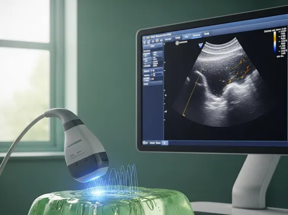
How High Resolution Micro Ultrasound Works
The fundamental principle behind High Resolution Micro Ultrasound is similar to that of conventional ultrasound, but with critical distinctions that enable its enhanced detail. An HRMUS system employs specialized transducers that emit very high-frequency sound waves into the body. These high-frequency waves have much shorter wavelengths compared to those used in standard ultrasound. As these shorter waves travel through tissues, they interact with various structures and reflect back to the transducer as echoes.
The transducer then receives these echoes, and a sophisticated computer system processes the timing, intensity, and frequency shifts of the returning sound waves to construct a detailed, real-time image. The trade-off for this superior resolution is a more limited penetration depth, making HRMUS particularly effective for imaging superficial organs and structures close to the body’s surface. This mechanism allows for the precise delineation of tissue layers, microvasculature, and cellular architecture, providing a level of detail previously unattainable with standard ultrasound.
Benefits and Applications of HRMUS Technology
The advent of High Resolution Micro Ultrasound has brought forth numerous advantages in clinical diagnostics. The primary benefits of high resolution ultrasound imaging include significantly improved diagnostic accuracy, enabling earlier detection of subtle abnormalities. This enhanced detail allows for better characterization of lesions, distinguishing between benign and malignant conditions with greater confidence. Furthermore, HRMUS is non-invasive, does not involve ionizing radiation, and provides real-time imaging, which is invaluable for guiding biopsies and other interventional procedures with precision.
The diverse applications of micro ultrasound technology are expanding across several medical fields. It has shown particular promise in:
- Prostate Imaging: For the detection and targeted biopsy of prostate cancer, offering superior visualization of suspicious lesions within the prostate gland.
- Dermatology: For detailed assessment of skin lesions, including melanoma and non-melanoma skin cancers, as well as inflammatory skin conditions.
- Vascular Imaging: To visualize superficial blood vessels and assess microvascular flow, aiding in the diagnosis of various vascular conditions.
- Musculoskeletal Imaging: For high-resolution evaluation of tendons, ligaments, and peripheral nerves, detecting subtle injuries or inflammatory changes.
- Small Parts Imaging: Enhancing the diagnostic capabilities for organs such as the thyroid, testes, and breast, by providing clearer images of small nodules or cysts.
These applications underscore HRMUS’s role as a valuable tool for clinicians seeking to improve patient outcomes through more precise and timely diagnoses. For instance, in prostate cancer detection, HRMUS has been shown to improve the detection rate of clinically significant cancers compared to conventional ultrasound-guided biopsies, as highlighted by studies in urological oncology.