Heterogeneously Dense Breast Tissue
Breast density is a classification based on the amount of fibrous and glandular tissue compared to fatty tissue in the breast. Understanding your breast density, particularly if you have heterogeneously dense breast tissue, is crucial for personalized breast cancer screening and risk assessment.
Key Takeaways
- Heterogeneously Dense Breast Tissue indicates a significant amount of glandular and fibrous tissue, making it harder to detect abnormalities on mammograms.
- This tissue type is associated with a slightly increased risk of developing breast cancer.
- Mammograms can be less effective in detecting tumors in dense breasts due to the masking effect of the dense tissue.
- Supplemental screening methods, such as ultrasound or MRI, may be recommended in addition to mammography for individuals with dense breasts.
- Regular consultation with a healthcare provider is essential to determine the most appropriate screening regimen based on individual risk factors.
What is Heterogeneously Dense Breast Tissue?
Heterogeneously Dense Breast Tissue refers to a common finding on mammograms where there are areas of both fatty tissue and non-fatty glandular and fibrous tissue. This classification means that while some fat is present, there are also regions of density that could obscure small masses on a mammogram. The presence of this tissue type is not abnormal or a disease in itself, but it is an important factor in breast cancer screening.
The heterogeneously dense breasts meaning indicates that the breast tissue is not uniformly dense, but rather a mix of densities. This differs from extremely dense breasts, which have very little fatty tissue, and fatty breasts, which consist almost entirely of fat. Breast density is categorized using the Breast Imaging Reporting and Data System (BI-RADS) by radiologists, ranging from A (almost entirely fatty) to D (extremely dense). Heterogeneously dense tissue falls into category C.
| BI-RADS Category | Description | Implication for Mammography |
|---|---|---|
| A | Almost entirely fatty | Easiest to detect abnormalities. |
| B | Scattered areas of fibroglandular density | Some dense tissue, but still good visibility. |
| C | Heterogeneously dense | Areas of non-dense tissue, but dense areas may obscure small masses. |
| D | Extremely dense | Most difficult to detect abnormalities; significantly reduced sensitivity. |
Risks and Implications of Heterogeneously Dense Breasts
Having heterogeneously dense breast tissue carries two primary implications for breast health. Firstly, dense breast tissue can make it more challenging for radiologists to detect breast cancer on a mammogram. Both dense tissue and cancerous tumors appear white on a mammogram, creating a “masking effect” that can hide potential abnormalities. This means that small cancers might be missed, leading to a delayed diagnosis.
Secondly, research indicates that women with dense breasts have a slightly higher risk of developing breast cancer compared to women with fatty breasts. While the exact mechanism is not fully understood, it is believed that the increased amount of glandular and fibrous tissue itself may contribute to this elevated risk. According to the American Cancer Society, women with heterogeneously dense breasts have a 1.2 to 2 times higher risk of breast cancer than women with average breast density. This increased risk, combined with the masking effect, underscores the importance of understanding one’s breast density.
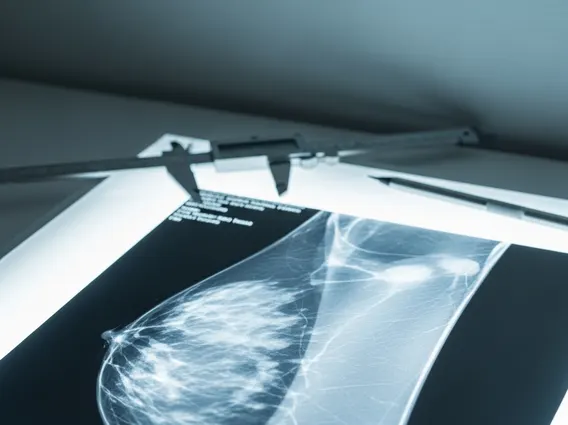
Mammogram Findings and Management of Dense Breast Tissue
When mammogram results heterogeneously dense are reported, it signifies that standard mammography alone may not be sufficient for comprehensive screening. Due to the reduced sensitivity of mammograms in dense breasts, healthcare providers often recommend supplemental screening methods to improve cancer detection rates. These additional screenings can help identify cancers that might be obscured by dense tissue.
Common supplemental screening options include:
- Breast Ultrasound: This non-invasive imaging technique uses sound waves to create images of breast tissue. It is particularly effective at distinguishing between solid masses (which could be tumors) and fluid-filled cysts, and it does not use radiation.
- Breast MRI (Magnetic Resonance Imaging): MRI uses powerful magnets and radio waves to produce detailed images of the breast. It is highly sensitive for detecting breast cancer in dense breasts but is typically reserved for women with a higher lifetime risk of breast cancer due to its cost and potential for false positives.
- 3D Mammography (Tomosynthesis): While still a mammogram, 3D mammography takes multiple images from different angles, creating a three-dimensional view of the breast. This can help radiologists see through dense tissue layers more effectively and reduce false alarms.
It is important for individuals to discuss their breast density and overall breast cancer risk factors with their doctor. Together, they can determine the most appropriate and personalized screening plan, which may include a combination of mammography and supplemental imaging, to ensure the best possible outcomes for early detection.