
Eklund Displacement Views
Eklund displacement views are a specialized mammographic technique designed to improve breast tissue visualization in patients with implants, ensuring that more of the natural breast tissue is captured and evaluated accurately.
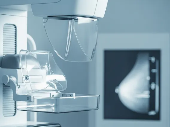
Key Takeaways
- Eklund displacement views push the implant posteriorly so more native breast tissue is imaged during mammography.
- This technique significantly increases the amount of breast tissue visualized compared to standard implant-present views.
- It is the recommended approach for breast cancer screening in patients with augmentation implants.
- Proper radiographer positioning is essential for obtaining diagnostic-quality images with this technique.
- Eklund views are used alongside standard implant-displaced mammograms for comprehensive evaluation.
How Eklund Displacement Views Improve Breast Imaging with Implants
The Eklund technique addresses a fundamental challenge in breast imaging: implants obstruct a substantial portion of breast tissue on conventional mammographic views. By manually displacing the implant posteriorly against the chest wall and pulling the native breast tissue forward over the implant, radiographers can compress and image significantly more glandular tissue than would otherwise be possible.
Research indicates that standard implant-present (IP) views capture only about 50–60% of breast tissue, whereas Eklund displacement views can increase visualization to approximately 80–85% of the total breast parenchyma. This improvement in tissue coverage is clinically meaningful, as obscured tissue represents areas where early lesions could go undetected. The technique effectively separates the implant from the tissue being compressed, reducing the artifact overlap that limits diagnostic confidence on IP views.
| View Type | Implant Position | Tissue Captured | Compression Quality |
|---|---|---|---|
| Implant-Present (IP) | Included in field | ~50–60% | Limited by implant |
| Eklund Displacement | Displaced posteriorly | ~80–85% | Improved glandular compression |
Eklund Displacement Views in Breast Cancer Screening: Clinical Role
Eklund views breast cancer screening protocols typically require four additional projections — mediolateral oblique (MLO) and craniocaudal (CC) displacement views for each breast — taken alongside the standard implant-present views. This eight-image protocol is endorsed by major radiology organizations to ensure thorough evaluation in augmented patients.
The clinical importance of this approach is underscored by the fact that breast implants are common among women of screening age. According to the American Society of Plastic Surgeons, breast augmentation consistently ranks among the most performed cosmetic procedures in the United States, meaning a substantial portion of the screening population has implants. Without displacement views, mammographic sensitivity is reduced, potentially delaying the detection of early-stage malignancies when treatment outcomes are most favorable.
Limitations do exist. Patients with subglandular implants, significant capsular contracture, or very large implants may have reduced benefit from the technique, as the implant cannot always be displaced sufficiently. In such cases, supplemental imaging modalities such as ultrasound or MRI may be recommended by the radiologist to complement mammographic findings.
Performing the Eklund Technique: Positioning and Mammogram Protocol
Correct execution of the Eklund technique breast implant mammogram positioning is critical to obtaining diagnostic images. The radiographer places both hands on the breast, using the fingers to firmly push the implant back toward the chest wall while simultaneously pulling the overlying glandular tissue anteriorly onto the image receptor. Once the implant is displaced, compression is applied to the native tissue only.
Key steps in performing this protocol include:
- Confirming implant type and placement (subglandular vs. submuscular) before beginning, as submuscular implants are generally easier to displace.
- Communicating clearly with the patient to minimize discomfort and promote cooperation during repositioning.
- Applying adequate but careful compression to maximize tissue spread without causing pain or implant stress.
Submuscular implants tend to respond more favorably to this technique because the pectoralis muscle provides a natural posterior anchor. Radiographers should document implant characteristics and any positioning limitations in the imaging report to assist radiologist interpretation. Adequate training and experience are essential, as improper technique can yield suboptimal views that reduce diagnostic accuracy.



















