Diagnosis, Screening, and Early Detection of Squamous Cell Carcinoma
Squamous cell carcinoma (SCC) is a common form of skin cancer that begins in the squamous cells of the skin’s outer layer. This article focuses on how SCC is recognized, who should have their skin checked, and which squamous cell carcinoma diagnosis methods confirm the disease.
Key Takeaways
- A persistent scaly patch, a sore that will not heal, or a firm, raised growth on sun-exposed skin is a common reason to have a lesion checked.
- Regular skin checks matter most for people who have had a lot of cumulative sun exposure, have fair skin, have battled skin cancer themselves or have a close relative who has, or have a suppressed immune system.
- A squamous cell carcinoma diagnosis is confirmed only through a skin biopsy — there is no blood test or imaging study that replaces it.
- Cutaneous SCC (on the skin) and the rarer non-cutaneous SCC (on mucous membranes) are found through different diagnostic paths.
- Catching SCC before it has had the chance to grow deeper or reach other sites generally translates into a more straightforward path through diagnosis and treatment.
Recognizing Early Signs of Squamous Cell Carcinoma
Noticing the earliest changes in the skin is what usually starts the diagnostic process for squamous cell carcinoma. SCC develops most often on skin that gets regular sun exposure, but it can also form on skin that has been injured, chronically inflamed, or previously treated with radiation, and less often on mucous membranes.
Common Visual Indicators
A full description of squamous cell carcinoma symptoms is covered separately; the indicators below are the ones that most often lead someone to seek a diagnostic evaluation:
- A scaly, red patch that bleeds or crusts and does not clear up.
- An open sore on the skin that does not heal.
- A firm, raised growth, which may have a central dip.
- A rough, scaly patch, sometimes on the lower lip, that keeps changing.
Because these patterns can look different from one person to the next, any new or changing skin lesion — not just ones matching this list — is worth having examined.
When to Seek Medical Advice
A clinician should look at any skin change that persists, grows, bleeds, or fails to heal within a few weeks. Waiting allows a tumor more time to grow or spread, which can make treatment more involved. Bringing a new or changing lesion to a clinician promptly is the practical starting point for early detection.
Screening Guidelines and Risk Factors for SCC
Screening means checking the skin before any symptoms are noticed, usually through a combination of self-exams and a clinical skin exam. Whether looking at large groups of otherwise healthy adults actually cuts the death toll from skin cancer remains unproven, which is why no fixed schedule is applied across the board; instead, clinicians generally tailor how often someone gets checked to that person’s own combination of risk factors.
Who Should Be Screened?
People with the following risk factors are the ones most likely to be advised to have their skin checked regularly:
- Extensive Sun Exposure: Long-term exposure to natural or artificial sunlight (including tanning beds) and a history of sunburns.
- Fair Complexion: Skin that burns easily or tans poorly, light-colored eyes, or red or blond hair — though people of any skin tone can develop SCC.
- Previous Skin Cancer: A prior diagnosis of basal cell carcinoma, SCC, or actinic keratosis — in yourself or in a parent or sibling — makes another occurrence more likely.
- Weakened Immune System: Organ transplant recipients and others on immunosuppressive medication have a significantly higher risk.
- Actinic Keratoses: These precancerous, scaly patches can progress to SCC over time.
Talking through these factors with a doctor helps determine how often skin checks make sense for a given person, rather than following one fixed schedule.
Personal Risk Assessment
A personal risk profile takes into account sun exposure history, skin type, family history, and immune status. Monthly self-exams help someone become familiar with their own skin so that new or changing spots stand out sooner. Combined with periodic professional checks for higher-risk individuals, this is the practical basis of squamous cell carcinoma screening.
Methods for Squamous Cell Carcinoma Diagnosis
Once a lesion looks suspicious, confirming squamous cell carcinoma requires removing a piece of it for examination under a microscope. No visual exam alone can make the diagnosis.
Biopsy Procedures Explained
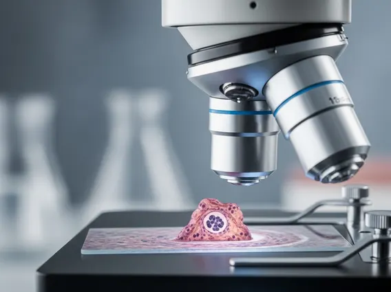
A skin biopsy is the only way to confirm SCC. A pathologist examines the tissue sample to check for cancer cells, identify the cell type, and estimate how deeply it has grown into the skin. Several biopsy techniques are used depending on the lesion:
- Shave Biopsy: A thin blade removes the top portion of the growth; this works well for lesions confined to the outer skin layers.
- Punch Biopsy: A small circular tool removes a deeper core of tissue, giving a fuller cross-section for evaluation.
- Excisional Biopsy: The entire visible growth is removed with a margin of surrounding tissue — this can serve as both diagnosis and initial treatment for a small, well-defined lesion.
- Incisional Biopsy: Only part of a larger growth is removed, often as a first step before planning further treatment.
Staging and Further Evaluation
After SCC is confirmed, further evaluation may follow for larger, deeper, or higher-risk tumors, since prognosis depends heavily on how far the cancer has spread as well as factors such as a weakened immune system or tobacco use. A clinician may check nearby lymph nodes by hand, order imaging — a CT scan, a chest film, a PET study, or an ultrasound of the area — and, in some cases, take a small sample from a lymph node for the pathologist to review. These findings feed into the staging process, which is covered in full on the dedicated staging page; here, the key point is that further testing is a diagnostic step used to plan the right treatment, not a routine part of every SCC diagnosis.
Types of Squamous Cell Carcinoma and Detection
Squamous cell carcinoma can develop in different locations, and where it forms changes how it is found.
Cutaneous vs. Non-Cutaneous SCC
Cutaneous SCC, the most common form, starts in the skin and is closely linked to ultraviolet exposure. It typically appears on sun-exposed areas such as the face, ears, neck, hands, and arms, and it is detected mainly through visual inspection followed by a skin biopsy.
Non-cutaneous SCC develops in mucous membranes, such as those lining the mouth and throat, rather than on the skin surface. Because these areas are harder to see and symptoms can be subtle, this form is often detected later. Smoking and infection with human papillomavirus (HPV) are established risk factors for squamous cell carcinoma of the mouth and throat, and diagnosis typically involves a direct examination plus a tissue biopsy of the affected area.
High-Risk Subtypes
Rather than a separate diagnostic category, certain circumstances are known to affect how squamous cell carcinoma behaves and is followed up: a weakened immune system, prior radiation to the area, or a lesion arising in a long-standing scar or area of chronic skin inflammation. Clinicians take these factors into account, alongside tumor size and depth found on biopsy, when deciding whether additional evaluation beyond the initial biopsy is warranted.
The Importance of Early Detection and Prognosis
Finding squamous cell carcinoma early is central to keeping treatment straightforward and protecting long-term health.
Impact on Treatment Outcomes
When SCC is identified while it is small and confined to the skin, treatment is generally more straightforward and recovery tends to go more smoothly; detailed outcome and survival figures by stage are addressed on the dedicated survival-rate page rather than here. If a lesion is left to grow, it can invade more deeply and, in some cases, reach nearby lymph nodes or other organs, which generally calls for more extensive treatment. This is why a prompt diagnostic workup after any suspicious change — rather than a wait-and-see approach — is the most useful step a patient can take.
Frequently Asked Questions
What are the most common signs of SCC?
The signs that most often prompt an evaluation are a persistent scaly or crusted patch, a sore that will not heal, and a firm, raised growth, usually on sun-exposed skin. Any new or changing skin lesion that lasts more than a few weeks is worth having a clinician examine.
Who is at highest risk for SCC and should be screened?
Higher risk falls on people with a lot of accumulated sun exposure, fair skin, an earlier skin cancer diagnosis of their own or in a close relative, or a compromised immune system. There is no single recommended screening interval for everyone; a doctor can help decide how often skin checks make sense based on individual risk factors.
How is SCC definitively diagnosed?
A biopsy is the only way to confirm squamous cell carcinoma. A small tissue sample is removed — using a shave, punch, incisional, or excisional technique depending on the lesion — and examined by a pathologist, who determines whether cancer cells are present and how deeply they extend.
Sources