Diagnosis, Screening, and Early Detection of Skin Cancer
Diagnosing skin cancer starts with a visual skin exam and, if a spot looks suspicious, a biopsy to confirm it under a microscope. This guide covers the skin cancer diagnosis methods clinicians use, what a professional screening visit involves, and how to check your own skin between appointments.
Key Takeaways
- A skin cancer diagnosis is confirmed by biopsy; a visual exam and dermatoscopy help a clinician decide when a biopsy is needed, but they cannot replace it.
- There is no single screening schedule recommended for everyone — current evidence is not sufficient to recommend routine population screening, so professional skin exams are usually discussed individually, especially for people with added risk factors.
- Learning the ABCDE warning signs of melanoma, and the different look of basal cell and squamous cell carcinoma, helps you recognize when a spot needs a doctor’s opinion.
- A monthly self-exam, done in the same systematic order each time, is a practical way to catch a new or changing spot between professional visits.
- Skin cancer found while still confined to the skin is generally easier to treat than skin cancer found after it has grown deeper or spread, which is why prompt evaluation of a changing spot matters.
Skin Cancer Diagnosis Methods
A skin cancer diagnosis usually begins with a visual examination by a doctor, who looks at the size, shape, color, and texture of a mole, sore, or other skin change. If something looks suspicious, the next step is typically a closer look with a dermatoscope, a biopsy, or both.
Types of Biopsies
A biopsy removes a sample of the abnormal skin so it can be examined under a microscope by a pathologist, and it is the definitive way to confirm a skin cancer diagnosis. The type of biopsy a doctor chooses mainly depends on where the growth is and how large it is:
- Shave biopsy: a sterile blade is used to shave off the top of the growth.
- Punch biopsy: a small, hollow circular tool removes a core of tissue, including some of the deeper skin layers.
- Excisional biopsy: the entire growth, along with a margin of surrounding skin, is cut out with a scalpel. Because it removes the whole lesion, this method also gives the pathologist a fuller picture of how deep an abnormal area extends, which matters most if the result turns out to be melanoma.
A second pathologist’s review can be requested if there is any uncertainty about the result.
Dermatoscopic Examination
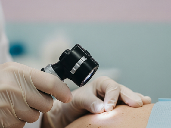
Dermatoscopy, also called dermoscopy, uses a handheld, illuminated magnifying device to examine a mole or lesion more closely than the naked eye allows. It helps a doctor look at pigment patterns, borders, and other structures that can point toward a benign mole or a concerning lesion, supporting the decision about whether a biopsy is needed. Even so, reading a pigmented lesion by sight is not always straightforward: studies comparing how different clinicians evaluate the same skin lesions have found real variability, which is one reason a biopsy remains the definitive step once a lesion looks suspicious.
Recognizing Early Skin Cancer Symptoms
Most skin cancer is first noticed as a change in the skin — a new growth, a sore that will not heal, or a mole that looks or behaves differently than before. Noticing a change like this is what leads someone to seek the evaluation described above; it is not a diagnosis on its own.
The ABCDE Checklist
Dermatologists often teach patients a simple letter-based checklist to describe mole changes worth flagging for melanoma, the least common but generally more aggressive type of skin cancer:
- Asymmetry: one half of the mole does not match the other.
- Border: the edges are jagged or poorly defined.
- Color: the color is uneven across the mole.
- Diameter: the mole is larger than a pea, though melanoma can sometimes be smaller.
- Evolving: the mole is changing in size, shape, or color, or has started to bleed or itch.
Any of these changes is a reason to have the spot looked at by a doctor.
Signs of Non-Melanoma Skin Cancers
Basal cell carcinoma often looks like a shiny bump, a flat pink or reddish patch, or a sore that bleeds, crusts, and does not fully heal. Squamous cell carcinoma often shows up as a firm bump that is red or close to the surrounding skin tone, or as a scaly, crusted spot that may bleed when irritated. Both usually develop on skin that gets regular sun exposure, such as the face, ears, neck, and hands, and either is worth having examined if it does not heal in the usual time frame.
Professional Skin Cancer Screening
Professional skin cancer screening means having a doctor examine your skin for spots that could be cancer. Unlike some other cancers, there is currently no single screening test or schedule recommended for the general population: the evidence available so far is not enough to say for certain whether routine full-body exams in people without symptoms reduce deaths from skin cancer, and screening also carries a chance of an unnecessary biopsy or of finding a lesion that would never have caused harm. Because of this, the decision to have periodic professional exams is usually made individually, based on a person’s own risk factors, rather than following one fixed rule for everyone.
Who Should Get Screened?
Anyone who notices a new or changing spot should have it evaluated, and some people may be advised by their doctor to have skin exams more often because they carry a higher risk of skin cancer. Factors that raise that risk include:
- Having had skin cancer before, or a close relative who has, including a melanoma diagnosis.
- A large number of moles, or moles with an atypical (irregular) appearance.
- Fair skin that burns or freckles easily, especially combined with sun exposure.
- A history of sunburns or frequent tanning.
- A weakened immune system, for example from medication taken after an organ transplant.
These factors add up rather than acting alone, so someone with several of them may want to talk to a doctor about a personal screening plan even without symptoms.
What to Expect During a Visit
A professional skin exam typically includes a review of your personal and family history and sun exposure, followed by a systematic, head-to-toe visual exam that covers spots people tend to overlook on their own, such as the scalp, the soles of the feet, the spaces between toes, and the back. The doctor may use a dermatoscope to look more closely at any spot of concern. Afterward, they will explain what they found and recommend next steps, such as a biopsy, if anything needs a closer look.
How to Perform a Skin Self-Exam
Checking your own skin regularly between professional visits helps you notice a new or changing spot early. Doing it the same way each time, roughly once a month, makes it easier to remember what your skin normally looks like.
Step-by-Step Guide
Use a full-length mirror, a hand mirror, and good lighting, ideally after a shower:
- Look at your face, ears, and neck, front and back.
- Part your hair with a comb to check your scalp, then use a hand mirror for the back of your neck and ears.
- Look over your palms and the top side of each hand, working between the fingers and under the nails, before moving up to the arms and underarms.
- In the full-length mirror, check your chest and abdomen, then use the hand mirror for your back.
- Sit down to check your thighs, shins, ankles, feet, and between your toes.
- Use a hand mirror to check your buttocks and genital area.
Look for anything that fits the ABCDE pattern described above, or a sore that is not healing, and report it to a doctor.
Key Areas to Check
A few spots are easy to overlook during a self-exam but can still develop skin cancer:
- Scalp: hidden by hair but still exposed to the sun.
- Behind the ears and neck: often missed because they are hard to see.
- Soles of the feet and between the toes: melanoma can occur here even without much sun exposure.
- Under the fingernails and toenails: a dark streak or bruise that does not grow out can be a sign of melanoma in the nail.
- Genital area and buttocks: not typically sun-exposed, but skin cancer can still develop there.
Why Early Detection Matters
Skin cancer caught while it is still confined to the skin is generally easier to treat, often with a single procedure that removes it completely, than skin cancer found after it has grown deeper or spread. Melanoma in particular spends time growing along the surface before it starts growing downward into deeper tissue, and according to the National Cancer Institute, more than 90% of melanomas can be spotted through simple visual inspection while still in that surface-level stage — before they grow into deeper tissue, which is when treatment becomes more complicated. Basal and squamous cell carcinomas are typically slower to spread, and finding them early usually still means a simpler procedure with less scarring. Across all three types, the general pattern holds: earlier detection and treatment are linked to better outcomes and fewer complications, which is the main reason self-exams and prompt evaluation of a new or changing spot are worth doing without delay.
Frequently Asked Questions
What are the different types of skin cancer?
The three main types are basal cell carcinoma, squamous cell carcinoma, and melanoma. Basal cell and squamous cell carcinoma are grouped together as keratinocyte carcinoma, are far more common overall, and usually appear on skin that gets regular sun exposure. Melanoma is less common but is generally the more aggressive of the three, so changes that fit the ABCDE pattern deserve prompt attention.
How often should I get a professional skin cancer screening?
There is no single recommended schedule for everyone: current evidence is not sufficient to say that routine screening benefits people without symptoms or extra risk factors. Many doctors still suggest periodic skin exams for people at higher risk, such as those with a personal or family history of skin cancer or many atypical moles. The best approach is to ask your own doctor what makes sense for your risk profile, and to report any new or changing spot as soon as you notice it rather than waiting for a scheduled visit.
Can skin cancer be prevented?
Not entirely, but the risk can be lowered with consistent sun protection: seeking shade during peak UV hours (10 a.m. to 4 p.m.), wearing protective clothing and a wide-brimmed hat, and applying a broad-spectrum sunscreen to exposed skin. Avoiding tanning beds is also recommended. Regular self-exams and, for some people, professional exams remain important because they catch what prevention alone cannot.