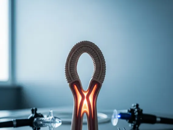
Carina Of Trachea
The carina of trachea is a small but anatomically critical structure located at the lower end of the windpipe, where the airway divides into the two main bronchi supplying the lungs. Its medical significance extends far beyond its modest size, playing a central role in airway assessment, disease staging, and bronchoscopic procedures.
Key Takeaways
- The carina of trachea is the ridge-like cartilaginous structure at the bifurcation of the trachea into the left and right main bronchi.
- It is located at the level of the sternal angle (angle of Louis), approximately at the T4–T5 vertebral level.
- Carina widening or distortion on imaging or bronchoscopy is a clinically significant sign of mediastinal pathology, including lymph node enlargement or tumor invasion.
- In lung cancer staging, tumor involvement of the carina is a key criterion that directly influences surgical planning and prognosis.
- Bronchoscopists routinely assess the carina to evaluate airway health, identify abnormalities, and guide diagnostic or therapeutic interventions.
Carina of Trachea: Anatomy, Location, and Structure
The carina of trachea refers to the sharp, keel-shaped cartilaginous ridge formed at the point where the trachea bifurcates into the right and left main bronchi. The term “carina” derives from the Latin word for keel, accurately describing its curved, projecting shape. This structure sits at the interior base of the trachea and serves as the dividing landmark between the single airway above and the dual bronchial tree below. The carina is lined with highly sensitive mucosa, making it one of the most reactive regions of the entire respiratory tract.
In terms of carina trachea location in the body, the structure lies at the level of the sternal angle — also called the angle of Louis — which corresponds to the intervertebral disc between the fourth and fifth thoracic vertebrae (T4–T5). In adults, this places the carina approximately 10 to 11 centimeters below the vocal cords, though precise positioning varies with height and respiratory phase. On a standard chest radiograph or computed tomography (CT) scan, the tracheal bifurcation angle, known as the subcarinal angle, typically measures between 55 and 65 degrees. An angle exceeding 70 degrees may indicate pathological enlargement of subcarinal lymph nodes or adjacent structures.
Structurally, the carina is composed of the same cartilaginous and fibromuscular tissue that forms the rest of the trachea. The right main bronchus departs at a more vertical angle (approximately 25 degrees from the midline), while the left main bronchus diverges at a wider angle (approximately 45 degrees). This anatomical asymmetry has practical consequences — foreign bodies, aspirated materials, and endotracheal tubes that are advanced too far tend to enter the right bronchus preferentially. Clinicians must understand this geometry when managing airway emergencies or placing airway devices.
Physiological Role and Medical Significance of the Carina of Trachea
The carina of trachea anatomy and function encompass both structural and physiological dimensions. Functionally, the carina acts as an aerodynamic divider, distributing inhaled air between the two lungs. The angle and shape of the carina influence airflow dynamics, affecting how particles, pathogens, and inhaled substances are distributed throughout the bronchial tree. Efficient airflow distribution is essential for adequate gas exchange and respiratory homeostasis.
One of the most clinically recognized functions of the carina is its role in triggering the cough reflex. The mucosal lining at the carina contains a dense concentration of cough receptors — rapidly adapting mechanoreceptors — that respond to mechanical stimulation, chemical irritants, and foreign material. Stimulation of these receptors initiates a powerful protective cough, which is why contact with the carina during endotracheal intubation or bronchoscopy frequently provokes vigorous coughing or bronchospasm. This reflex is a vital airway defense mechanism, preventing aspiration and clearing debris.
The carina of trachea medical significance explained in a clinical context centers on its use as a landmark for radiological and endoscopic assessment. Abnormalities at or near the carina — such as splaying, mucosal irregularity, erythema, or loss of the sharp carinal ridge — are recognized indicators of underlying disease. Enlargement of subcarinal lymph nodes, which lie directly beneath the carina, is a common finding in conditions including sarcoidosis, tuberculosis, lymphoma, and metastatic lung cancer. Radiologists and pulmonologists routinely evaluate the subcarinal angle and carinal morphology as part of a comprehensive thoracic assessment.
Carina Involvement in Lung Cancer Staging and Tumor Classification
The relationship between the carina of trachea and lung cancer staging is well-established within the TNM (Tumor, Node, Metastasis) classification system developed by the American Joint Committee on Cancer (AJCC) and the Union for International Cancer Control (UICC). In this framework, the proximity of a primary lung tumor to the carina is a defining criterion for T-stage classification. A tumor located less than 2 centimeters from the carina — but without direct invasion — is classified as T3, while a tumor that directly invades the carina itself is classified as T4, denoting locally advanced disease.
Carina trachea involvement in tumor staging carries profound prognostic implications. T4 tumors involving the carina are generally considered unresectable by standard surgical approaches, and their management typically involves multimodal strategies including concurrent chemoradiotherapy. According to data referenced in the AJCC Cancer Staging Manual (8th edition), patients with T4 disease have a significantly lower five-year survival rate compared to those with T3 or lower-stage tumors, underlining the importance of precise carinal assessment. Positron emission tomography-computed tomography (PET-CT) and endobronchial ultrasound (EBUS) are now standard tools for evaluating carinal involvement before treatment decisions are made.
Subcarinal lymph node involvement is separately staged under the nodal (N) component of TNM classification. Metastasis to subcarinal nodes — designated as station 7 in the International Association for the Study of Lung Cancer (IASLC) lymph node map — is classified as N2 disease when ipsilateral and N3 when contralateral, each carrying distinct management implications. Understanding the anatomical relationship between the carina and these nodal stations is therefore fundamental to accurate lung cancer staging and treatment planning.
| TNM T-Stage | Carinal Relationship | Clinical Implication |
|---|---|---|
| T2 | Tumor ≥ 2 cm from carina, no invasion | Potentially resectable; standard surgical evaluation |
| T3 | Tumor < 2 cm from carina, no direct invasion | Requires careful preoperative planning; may be resectable |
| T4 | Direct tumor invasion of the carina | Locally advanced; typically managed with multimodal therapy |
Clinical Relevance of the Carina in Bronchoscopy and Airway Procedures
The role of carina in bronchoscopy procedure is central to both diagnostic and therapeutic airway management. During flexible bronchoscopy, the carina is one of the first identifiable landmarks encountered after passing through the vocal cords and subglottic space. Bronchoscopists systematically inspect the carina for sharpness of its ridge, symmetry of the two main bronchi, mucosal color and texture, and the presence of any external compression or intrinsic lesions. A blunted, widened, or distorted carina is an important sign that prompts further investigation.
Endobronchial ultrasound-guided transbronchial needle aspiration (EBUS-TBNA) is a minimally invasive procedure that has transformed the sampling of subcarinal and paratracheal lymph nodes. Using a bronchoscope equipped with an ultrasound transducer, clinicians can visualize nodes immediately adjacent to the carina and obtain tissue samples under real-time imaging guidance, avoiding more invasive surgical mediastinoscopy in many cases. This technique has substantially improved the accuracy of lung cancer staging, with reported sensitivity rates exceeding 85% for mediastinal node sampling according to published systematic reviews in thoracic oncology literature.
Carinal Assessment During Intubation and Critical Care
During endotracheal intubation, the carina serves as a critical depth reference point. An endotracheal tube advanced beyond the carina will selectively intubate one bronchus — most often the right — resulting in single-lung ventilation and consequent hypoxia. Clinicians confirm correct tube placement using auscultation, end-tidal carbon dioxide monitoring, and chest radiography, with the tube tip ideally positioned 3 to 5 centimeters above the carina. In critically ill patients on mechanical ventilation, inadvertent right mainstem intubation is a recognized and preventable complication that reinforces the carina’s practical importance in daily clinical care.
Carinal Resection as a Surgical Technique
In select cases of centrally located lung tumors or primary tracheal tumors involving the carina, surgical resection of the carina itself — known as carinal resection — may be performed by thoracic surgeons. This technically demanding procedure involves removing the diseased carinal segment and reconstructing the airway by anastomosing the trachea to one or both bronchi. Patient selection, surgical expertise, and postoperative airway management are critical determinants of outcome. Carinal resection represents one of the most complex operations in thoracic surgery, reflecting both the anatomical centrality and functional indispensability of this structure.
Frequently Asked Questions
Where exactly is the carina of trachea located?
The carina of trachea is located at the lower end of the trachea where it divides into the right and left main bronchi. Anatomically, this corresponds to the level of the sternal angle (angle of Louis), at approximately the T4–T5 vertebral level. It lies within the mediastinum, surrounded by important structures including the aortic arch, pulmonary vessels, and mediastinal lymph nodes. In adults, it is positioned roughly 10 to 11 centimeters below the vocal cords, varying slightly with an individual’s body size and respiratory phase.
How does carina involvement affect lung cancer treatment options?
Direct invasion of the carina by a lung tumor elevates the tumor to T4 classification in the TNM staging system, which indicates locally advanced disease. This significantly limits surgical resectability and generally shifts treatment toward multimodal strategies such as concurrent chemotherapy and radiation therapy. In highly selected patients at specialized centers, carinal resection with airway reconstruction may be considered. Accurate staging through imaging and EBUS-guided biopsy is essential to determine whether the carina is truly involved before committing to a treatment plan.
Why is the carina sensitive and clinically important during bronchoscopy?
The carina contains a high density of cough receptors in its mucosal lining, making it extremely sensitive to mechanical or chemical stimulation. During bronchoscopy, contact with the carina commonly triggers coughing or bronchospasm, which clinicians anticipate and manage with appropriate sedation and topical anesthesia. Beyond its reflex sensitivity, the carina serves as a key anatomical landmark for assessing airway health, identifying signs of extrinsic compression or tumor invasion, and guiding the placement of instruments for subcarinal lymph node sampling.