Key Takeaways
- Renal Cell Carcinoma is the most prevalent form of kidney cancer, primarily affecting adults.
- Common renal cell carcinoma symptoms include blood in the urine, flank pain, and a palpable mass, though often it is asymptomatic in early stages.
- Risk factors for RCC include smoking, obesity, hypertension, and certain genetic conditions.
- Diagnosis typically involves imaging studies like CT scans and MRI, often followed by biopsy, to determine the extent and types of renal cell carcinoma.
- Treatment for renal cell carcinoma ranges from surgery (nephrectomy) to targeted therapies, immunotherapy, and radiation, with the choice depending on the stage and characteristics of the cancer.
Understanding Renal Cell Carcinoma
Renal Cell Carcinoma (RCC) is a malignancy that originates in the cells of the renal tubules, which are tiny tubes in the kidneys responsible for filtering waste from the blood. It represents approximately 90% of all kidney cancers and is more common in men than women, typically affecting individuals over the age of 50. Understanding what is renal cell carcinoma involves recognizing its cellular origin and its potential to spread if not detected and treated early.
What is RCC?
RCC develops when cells in the kidney’s lining grow uncontrollably, forming a tumor. These tumors can grow quite large before causing noticeable symptoms, which is why they are often discovered incidentally during imaging for other conditions. The kidneys are vital organs located on either side of the spine, below the rib cage, playing a crucial role in filtering blood, producing urine, and regulating blood pressure and red blood cell production. When RCC affects these functions, it can lead to various health complications.
According to the American Cancer Society, kidney cancer incidence rates have been slowly rising over the past few decades, emphasizing the importance of awareness and early detection strategies. While the exact reasons for this increase are complex, it highlights the need for continued research into its causes and effective treatments.
Key Types of Kidney Cancer
While Renal Cell Carcinoma is the overarching term, there are several distinct types of renal cell carcinoma, each with unique cellular characteristics and clinical behaviors. The most common types include:
- Clear Cell Renal Cell Carcinoma (ccRCC): This is the most prevalent type, accounting for 75-80% of all RCC cases. It originates from the proximal renal tubule and is characterized by cells that appear “clear” under a microscope due to their high lipid and glycogen content.
- Papillary Renal Cell Carcinoma (pRCC): Representing 10-15% of RCCs, pRCC forms finger-like projections (papillae) within the tumor. It is further divided into Type 1 and Type 2, with different genetic mutations and prognoses.
- Chromophobe Renal Cell Carcinoma: This less common type makes up about 5% of RCCs. It is characterized by cells with prominent cell membranes and pale, eosinophilic cytoplasm, and generally has a better prognosis than clear cell RCC.
- Collecting Duct Renal Cell Carcinoma: A rare and aggressive form, originating in the collecting ducts of the kidney.
- Unclassified Renal Cell Carcinoma: This category is used for tumors that do not fit into any of the recognized types or have features of multiple types.
Distinguishing between these types is crucial for accurate diagnosis and for guiding the most appropriate treatment for renal cell carcinoma, as their responses to therapies can differ significantly.
Recognizing Symptoms and Risk Factors
Early-stage Renal Cell Carcinoma often presents with no symptoms, making early detection challenging. However, as the tumor grows, it can lead to a range of signs and symptoms. Understanding these indicators and the associated risk factors is vital for prompt medical attention and diagnosis.
Common Symptoms of RCC
The classic triad of renal cell carcinoma symptoms includes blood in the urine, flank pain, and a palpable abdominal mass. However, this triad is only present in a small percentage of patients, usually indicating more advanced disease. More commonly, patients experience one or two of these, or more general symptoms. Common symptoms include:
- Hematuria: Blood in the urine, which can be visible (gross hematuria) or microscopic (detected only under a microscope). This is often the first and most common symptom.
- Flank Pain: Persistent pain or pressure in the side or back, just below the ribs, which is not related to injury.
- Abdominal Mass: A lump or mass that can be felt in the abdomen or side.
- Fatigue: Persistent tiredness and lack of energy, often due to anemia caused by chronic blood loss or the cancer itself.
- Unexplained Weight Loss: Significant loss of weight without changes in diet or exercise.
- Fever: Persistent low-grade fever not caused by an infection.
- Anemia: A decrease in red blood cells, leading to paleness and weakness.
- Swelling in Ankles or Legs: Can occur if the tumor affects blood flow.
It is important to note that these symptoms can also be caused by other, less serious conditions. Therefore, any new or persistent symptoms warrant a visit to a healthcare professional for proper evaluation.
Causes and Risk Factors
While the exact causes of renal cell carcinoma are not fully understood, several risk factors have been identified that increase an individual’s likelihood of developing the disease. These factors often involve a combination of genetic predispositions and environmental exposures:
- Smoking: Tobacco use significantly increases the risk of RCC. The risk increases with the amount and duration of smoking.
- Obesity: Being overweight or obese is a well-established risk factor, possibly due to hormonal changes associated with excess body fat.
- High Blood Pressure (Hypertension): Individuals with high blood pressure, particularly those on certain medications, have an increased risk.
- Advanced Kidney Disease and Dialysis: Long-term dialysis for chronic kidney failure can increase the risk of developing cystic kidney disease, which can sometimes lead to RCC.
- Certain Genetic Syndromes: Inherited conditions such as von Hippel-Lindau (VHL) disease, Birt-Hogg-Dubé syndrome, hereditary papillary renal cell carcinoma, and hereditary leiomyomatosis and renal cell carcinoma are strongly linked to an increased risk of RCC.
- Exposure to Certain Substances: Occupational exposure to substances like cadmium, asbestos, and trichloroethylene has been associated with a higher risk.
- Family History: Having a close relative (parent, sibling, child) with kidney cancer can slightly increase an individual’s risk.
While some risk factors like genetics are unchangeable, lifestyle modifications such as quitting smoking, maintaining a healthy weight, and managing blood pressure can help reduce the risk of developing Renal Cell Carcinoma.
Diagnosing Renal Cell Carcinoma
The accurate and timely renal cell carcinoma diagnosis is crucial for effective treatment planning. Due to the often asymptomatic nature of early RCC, many cases are discovered incidentally during imaging tests performed for other health concerns. Once symptoms arise or a suspicious mass is found, a series of diagnostic methods are employed to confirm the presence of cancer and determine its extent.
Diagnostic Methods and Staging
The diagnostic process for Renal Cell Carcinoma typically involves a combination of medical history review, physical examination, imaging studies, and sometimes a biopsy. Staging is a critical part of diagnosis, as it describes the size of the tumor and whether it has spread, guiding treatment decisions and influencing the renal cell carcinoma prognosis.
Key diagnostic methods include:
- Imaging Tests:
- Ultrasound: Often the first imaging test, it can detect kidney masses and distinguish between solid tumors and fluid-filled cysts.
- Computed Tomography (CT) Scan: A detailed imaging technique that provides cross-sectional images of the kidneys and surrounding structures, helping to identify the size, location, and spread of the tumor.
- Magnetic Resonance Imaging (MRI): Used when CT scans are inconclusive, or if a patient cannot receive CT contrast dye. It offers excellent soft tissue contrast.
- Positron Emission Tomography (PET) Scan: Less commonly used for initial diagnosis of RCC, but can be helpful in detecting distant metastases.
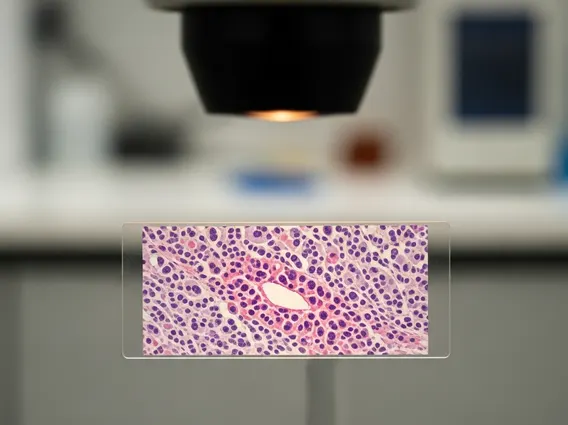
- Biopsy: While imaging can strongly suggest RCC, a biopsy (removing a small tissue sample for microscopic examination) is sometimes performed to confirm the diagnosis and determine the specific types of renal cell carcinoma. However, it’s not always necessary if imaging is highly suggestive and surgery is planned.
- Blood and Urine Tests: These tests assess kidney function, check for anemia, and look for other markers that might indicate cancer or its effects on the body.
Staging: Once RCC is diagnosed, it is staged using the TNM (Tumor, Node, Metastasis) system, which describes:
| Component | Description |
|---|---|
| T (Tumor) | Indicates the size and extent of the primary tumor within the kidney and whether it has grown into nearby tissues. |
| N (Nodes) | Determines if the cancer has spread to nearby lymph nodes. |
| M (Metastasis) | Indicates whether the cancer has spread to distant parts of the body (e.g., lungs, bones, liver, brain). |
This staging information is crucial for physicians to develop an individualized treatment for renal cell carcinoma plan and to provide an accurate outlook.
Treatment Approaches and Prognosis
The treatment for renal cell carcinoma is highly individualized, depending on the cancer’s stage, the specific types of renal cell carcinoma, the patient’s overall health, and personal preferences. Advances in medical science have significantly expanded the range of available therapies, offering improved outcomes for many patients.
Current Treatment Options
Treatment strategies for Renal Cell Carcinoma aim to remove or destroy the cancer cells, control tumor growth, and manage symptoms. The primary treatment modalities include:
- Surgery: This is the most common and often curative treatment for localized RCC.
- Radical Nephrectomy: Removal of the entire kidney, along with the adrenal gland and surrounding fatty tissue and lymph nodes.
- Partial Nephrectomy: Removal of only the part of the kidney containing the tumor, preserving healthy kidney tissue. This is often preferred for smaller tumors or when the patient has only one kidney.
- Ablation Therapies: For smaller tumors or patients who cannot undergo surgery, these minimally invasive techniques destroy cancer cells using extreme temperatures.
- Radiofrequency Ablation (RFA): Uses heat generated by radio waves.
- Cryoablation: Uses extreme cold to freeze and destroy tumor cells.
- Targeted Therapy: These drugs specifically target pathways involved in cancer cell growth and blood vessel formation (angiogenesis) that tumors need to survive. Examples include tyrosine kinase inhibitors (TKIs) and mTOR inhibitors.
- Immunotherapy: These treatments boost the body’s own immune system to recognize and destroy cancer cells. Checkpoint inhibitors, such as PD-1/PD-L1 inhibitors, have shown significant success in advanced RCC.
- Radiation Therapy: While RCC is generally resistant to traditional radiation, it can be used to manage symptoms like pain from bone metastases or to treat brain metastases. Stereotactic body radiation therapy (SBRT) is a more precise form that delivers high doses to the tumor with minimal damage to surrounding healthy tissue.
- Chemotherapy: Traditional chemotherapy is generally not very effective against RCC and is rarely used as a primary treatment.
Often, a combination of these therapies is used, especially for advanced or metastatic disease, to achieve the best possible outcome.
Outlook and Follow-up
The renal cell carcinoma prognosis varies significantly depending on several factors, including the stage of the cancer at diagnosis, the specific type of RCC, the patient’s overall health, and their response to treatment. Early detection and localized disease generally lead to a much better prognosis.
According to the National Cancer Institute’s SEER program data (2013-2019), the overall 5-year relative survival rate for kidney and renal pelvis cancer is approximately 77.6%. However, this rate can be much higher for localized disease (93.3%) and significantly lower for metastatic disease (15.8%). These statistics highlight the critical importance of early diagnosis.
After initial treatment, regular follow-up care is essential. This typically involves periodic physical examinations, blood tests, and imaging scans (CT, MRI) to monitor for recurrence or the development of new tumors. The frequency and type of follow-up depend on the stage of the original cancer and the chosen treatment. Long-term surveillance helps ensure that any potential issues are identified and addressed promptly, contributing to a better long-term outlook for individuals treated for Renal Cell Carcinoma.
It is important for patients to discuss their specific prognosis with their healthcare team, as individual circumstances can vary widely.
Renal Cell Carcinoma FAQs
Renal Cell Carcinoma most commonly affects individuals in their 50s, 60s, and 70s, with the average age at diagnosis being around 64. While it can occur at any age, including in younger adults and children, it is relatively rare in these populations. The incidence rate tends to increase with age, making it primarily a disease of older adults. This demographic trend is important for understanding population-level risk and screening considerations.
Yes, Renal Cell Carcinoma can be cured, especially when it is diagnosed at an early stage and remains localized to the kidney. For localized tumors, surgical removal (nephrectomy) is often curative. Even in some cases of advanced or metastatic RCC, significant progress has been made with targeted therapies and immunotherapy, offering long-term disease control and improved survival for many patients. The likelihood of cure depends heavily on the stage and characteristics of the cancer at diagnosis.
Several lifestyle changes can help reduce the risk of developing Renal Cell Carcinoma. Quitting smoking is paramount, as tobacco use is a major risk factor. Maintaining a healthy weight through balanced diet and regular exercise can mitigate the risk associated with obesity. Managing high blood pressure effectively, either through lifestyle modifications or medication, is also crucial. Limiting exposure to certain industrial chemicals like cadmium and trichloroethylene, where possible, can further contribute to risk reduction.