

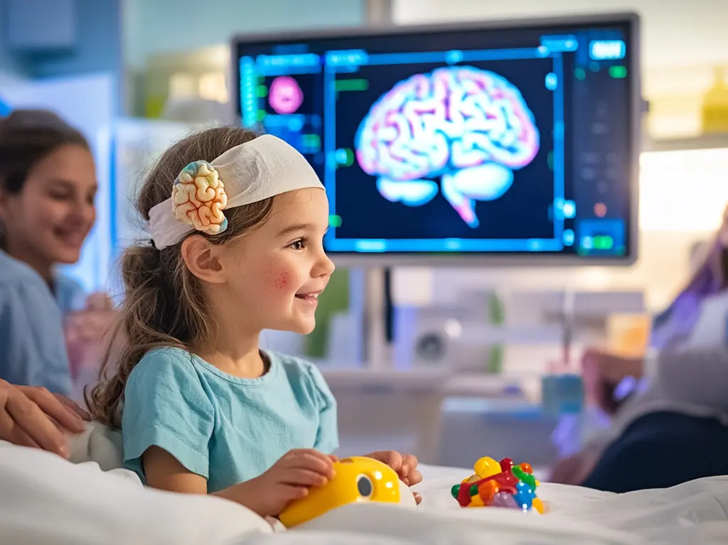
Atypical teratoid rhabdoid tumor is one of the rarest and most aggressive brain tumors diagnosed in early childhood, requiring urgent medical attention from the moment of suspicion. Understanding the nature of this condition, from its biological origins to available therapies, can help families and caregivers make informed decisions alongside their medical team.
Key Takeaways
- ATRT cancer is a rare, high-grade brain and spinal cord tumor that predominantly affects children under three years of age.
- The condition is strongly linked to mutations or deletions in the SMARCB1 (INI1) gene, though most cases arise sporadically without a family history.
- Symptoms vary by tumor location but often include vomiting, lethargy, difficulty walking, and changes in eye movement or head size in infants.
- Diagnosis relies on MRI imaging, cerebrospinal fluid analysis, and molecular pathology confirmation, while survival rates remain significantly lower than for many other pediatric brain tumors.
- Treatment typically combines surgery, radiation therapy, and intensive chemotherapy, with emerging clinical trials offering additional hope for eligible patients.
What Is ATRT Cancer and Who Does It Affect?
Atypical teratoid rhabdoid tumor (ATRT) is a fast-growing, malignant central nervous system tumor that arises most commonly in the brain but can also develop along the spinal cord. It is classified as a World Health Organization (WHO) Grade 4 tumor, meaning it is among the highest-grade and most aggressive neoplasms recognized in pediatric neuro-oncology. Despite its rarity, ATRT accounts for approximately 1–2% of all pediatric brain tumors and roughly 10–20% of brain tumors diagnosed in children under three years of age, according to data published in peer-reviewed pediatric oncology literature.
The tumor predominantly affects infants and very young children. The median age at diagnosis is around 17 months, and the vast majority of cases occur before a child’s third birthday. While ATRT can technically arise at any age, presentation in older children or adults is exceptionally uncommon. The tumor does not show a strong sex predilection, though some studies suggest a slight male predominance. Because it occurs during such a critical window of neurological development, it presents unique clinical and treatment challenges that differ substantially from brain tumors in older patients.
ATRT tumors can appear in several regions of the central nervous system. The posterior fossa — which houses the cerebellum and brainstem — is the most frequent site, accounting for roughly half of all cases. Supratentorial locations (the cerebral hemispheres) and the spinal cord represent the remaining cases. Tumor location plays a significant role in determining both the presenting symptoms a child experiences and the feasibility of surgical removal, making early and accurate identification of the tumor’s anatomical site essential to planning care.
Causes, Risk Factors, and Symptoms of Atypical Teratoid Rhabdoid Tumor
The defining molecular characteristic of ATRT cancer is the loss of function of the SMARCB1 gene, also known as INI1 or hSNF5, located on chromosome 22. This gene encodes a protein that is part of the SWI/SNF chromatin-remodeling complex, which plays a fundamental role in regulating gene expression and suppressing tumor growth. When SMARCB1 is mutated, deleted, or otherwise inactivated, cells lose a critical brake on proliferation, allowing malignant tumor formation. In a minority of cases, a related gene called SMARCA4 is implicated instead.
Most occurrences of ATRT are sporadic, meaning they arise from new (de novo) genetic mutations with no inherited family history. However, approximately 25–35% of cases are associated with rhabdoid tumor predisposition syndrome (RTPS), a hereditary condition in which a germline mutation in SMARCB1 or SMARCA4 is inherited from a parent. Families with a history of rhabdoid tumors — whether in the brain, kidneys, or soft tissues — may benefit from genetic counseling to assess inherited risk. Beyond this genetic predisposition, no confirmed environmental, dietary, or lifestyle risk factors have been identified as contributing to ATRT development.
The atypical teratoid rhabdoid tumor symptoms in children vary depending on where in the central nervous system the tumor is located and how rapidly it is growing. Common presentations include:
- Persistent or projectile vomiting, particularly in the morning
- Lethargy, irritability, or marked changes in behavior
- Poor feeding or failure to thrive in infants
- Abnormal or rapid increase in head circumference (macrocephaly) in very young infants
- Difficulty with balance, coordination, or walking (ataxia)
- Abnormal eye movements, including nystagmus or gaze palsy
- Facial weakness or cranial nerve deficits
- Seizures, in some cases
Because many of these signs can overlap with benign conditions common in early infancy, diagnosis is frequently delayed. Clinicians and caregivers should be alert to combinations of neurological symptoms — especially when accompanied by rapid head growth or persistent morning vomiting — that warrant prompt imaging evaluation.
ATRT Cancer Diagnosis and Survival Rate
Diagnosing ATRT requires a combination of advanced neuroimaging, tissue biopsy, and molecular testing. Magnetic resonance imaging (MRI) of the brain and entire spine is the primary imaging modality and can reveal tumor size, location, and evidence of dissemination through the cerebrospinal fluid (CSF) pathways. Tumors often appear as large, heterogeneous masses with areas of hemorrhage, necrosis, or calcification on imaging. Full spinal staging is essential because ATRT has a notable tendency to spread via CSF, occurring in roughly 20–35% of patients at the time of diagnosis.
Definitive diagnosis requires surgical tissue sampling, after which neuropathologists perform immunohistochemistry (IHC) to confirm the loss of INI1 (SMARCB1) protein expression — the hallmark finding that distinguishes ATRT from other embryonal brain tumors such as medulloblastoma. Molecular testing, including next-generation sequencing, further characterizes the tumor and can help guide treatment decisions. Lumbar puncture to examine CSF for tumor cells is also performed as part of staging, provided it is safe to do so given the child’s intracranial pressure.
The ATRT cancer diagnosis and survival rate data reflect the significant challenges this tumor presents. Overall five-year survival rates have historically been reported between 20% and 40%, though outcomes vary considerably based on age at diagnosis, extent of surgical resection, presence of metastatic disease, and access to specialized treatment centers. Children older than three years at diagnosis and those with localized (non-metastatic) tumors tend to have relatively better outcomes. Research from institutions participating in international pediatric oncology consortia has shown gradual improvement in survival over recent decades as treatment protocols have intensified, though ATRT remains one of the most difficult pediatric brain tumors to treat successfully.
| Factor | Associated Outcome |
|---|---|
| Age < 3 years at diagnosis | Generally poorer prognosis |
| Metastatic disease at diagnosis | Significantly reduced survival |
| Gross total surgical resection | Associated with better outcomes |
| Localized (non-metastatic) tumor | Improved survival rates |
| Germline SMARCB1 mutation (RTPS) | May indicate higher risk of recurrence |
Treatment Options for ATRT Brain Tumors in Young Children
Managing ATRT requires a multidisciplinary team that includes pediatric neurosurgeons, neuro-oncologists, radiation oncologists, and specialist nurses working together to design an individualized treatment plan. Because of the tumor’s aggressive nature and the young age of most patients, treatment must balance maximizing efficacy against minimizing long-term neurodevelopmental harm. No single universally accepted standard protocol exists worldwide, but most current approaches incorporate three main modalities: surgery, chemotherapy, and radiation therapy.
Surgical resection is typically the first step, with the goal of achieving gross total removal of the tumor whenever safely possible. Studies consistently demonstrate that more complete resection correlates with better survival outcomes. However, the proximity of many ATRT tumors to critical brainstem and cerebellar structures means that total removal is not always achievable without unacceptable neurological risk. Following surgery, systemic chemotherapy is initiated promptly. Regimens often include combinations of agents such as vincristine, cisplatin, cyclophosphamide, etoposide, and high-dose methotrexate, sometimes alongside intrathecal (directly into the CSF) chemotherapy to address potential microscopic spread throughout the spinal axis.
Radiation therapy is a highly effective component of ATRT treatment but presents particular concerns in very young children, as craniospinal or focal brain irradiation can cause significant long-term cognitive and endocrine effects in the developing brain. For children under three years of age, radiation is often deferred or modified in intensity, relying more heavily on chemotherapy regimens in the interim. For older children, focal or craniospinal radiation is incorporated into the treatment plan and has been associated with improved local tumor control. Proton beam therapy, where available, is increasingly preferred over conventional photon radiation because it delivers a more precise dose with less exposure to surrounding healthy brain tissue.
The atypical teratoid rhabdoid tumor treatment options landscape is evolving rapidly, with active research focusing on molecularly targeted agents and immunotherapies. Because the loss of SMARCB1 disrupts the SWI/SNF complex, researchers are investigating EZH2 inhibitors and other epigenetic therapies designed to restore normal gene regulation in tumor cells. Clinical trials sponsored by the Children’s Oncology Group (COG), the Society for Neuro-Oncology (SNO), and international consortia are enrolling patients to evaluate these novel approaches. Families are strongly encouraged to discuss clinical trial eligibility with their oncology team, as participation may provide access to promising therapies not yet available through standard care.
Supportive care is an equally integral component of the overall treatment strategy. Rehabilitation services — including physical therapy, occupational therapy, speech therapy, and neuropsychological support — are initiated early and continued throughout and after active treatment to preserve and restore as much function as possible. Palliative care specialists also play a role in managing pain, treatment side effects, and the broader emotional needs of the child and family throughout the care journey.
Frequently Asked Questions
Is ATRT cancer hereditary?
Most ATRT cases arise from spontaneous genetic mutations with no family history, making them sporadic in nature. However, approximately 25–35% of cases are linked to rhabdoid tumor predisposition syndrome, caused by an inherited germline mutation in the SMARCB1 or SMARCA4 gene. Families with a known history of rhabdoid tumors in any body site should consult a genetic counselor to understand their specific risk and discuss appropriate surveillance or testing options for other family members.
Can ATRT cancer recur after treatment?
Recurrence is a significant concern with ATRT, as the tumor is highly aggressive and can return locally or spread through the CSF pathways even after initial treatment achieves remission. Recurrent ATRT carries a particularly poor prognosis, and treatment options at relapse are more limited. Ongoing monitoring through regular MRI scans and clinical follow-up is essential. Some patients with recurrent disease may be eligible for experimental therapies or salvage chemotherapy regimens through clinical trials at specialized pediatric cancer centers.
Are there long-term effects of ATRT treatment in young children?
Because treatment occurs during a critical window of brain development, survivors of ATRT may experience long-term neurocognitive effects, including difficulties with memory, attention, learning, and executive function — particularly if radiation therapy was used. Endocrine disorders, hearing loss from platinum-based chemotherapy, and physical developmental delays are also reported. Comprehensive long-term follow-up through a survivors’ program is essential to monitor for late effects and provide appropriate rehabilitative, educational, and psychological support as children grow.



















