Nevus
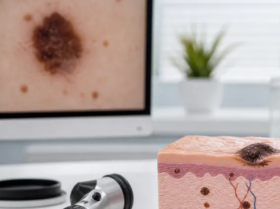
A nevus, commonly known as a mole, is a benign growth on the skin that can vary widely in appearance. Understanding these common skin lesions is important for general health awareness and for identifying potential concerns.
Key Takeaways
- A Nevus is a common, benign skin lesion resulting from the proliferation of melanocytes.
- Nevi come in various forms, including junctional, compound, dermal, and atypical types, each with distinct characteristics.
- While most nevi are harmless, it’s crucial to monitor them for changes in size, shape, color, or texture, which could indicate a need for medical evaluation.
- Diagnosis typically involves visual inspection by a dermatologist, sometimes followed by dermoscopy or biopsy.
- Regular self-skin exams and professional check-ups are vital for early detection of any suspicious lesions.
What is Nevus: Understanding This Common Skin Condition
A Nevus refers to a common, benign skin lesion characterized by the proliferation of melanocytes, the pigment-producing cells in the skin. These growths are extremely common, with most adults having between 10 and 40 nevi. They can appear anywhere on the body, from the scalp to the soles of the feet, and vary significantly in size, shape, and color. This nevus skin condition is generally harmless, but understanding its nature is crucial for distinguishing between normal moles and those that might require medical attention.
Nevi typically develop during childhood and adolescence, though new ones can appear throughout life. Their development is influenced by genetics and sun exposure. While most nevi are stable, some may change over time, and a small percentage can evolve into melanoma, a serious form of skin cancer. Therefore, regular monitoring and professional evaluation of any changing or suspicious lesions are essential for early detection and intervention.
Types of Nevi and Their Characteristics
The classification of nevi is based on their histological features and clinical appearance, leading to various types of nevus and their characteristics. Understanding these distinctions helps in assessing their benign nature or potential for concern. Here are some of the most common types:
- Junctional Nevus: These nevi are typically flat or slightly raised and uniformly brown or black. The melanocytes are located at the junction between the epidermis and dermis.
- Compound Nevus: Characterized by melanocytes present in both the epidermis and the dermis, compound nevi are usually slightly raised, dome-shaped, and may be pigmented or skin-colored. They often have a smooth surface.
- Intradermal Nevus: In this type, melanocytes are found exclusively within the dermis. Intradermal nevi are often raised, dome-shaped, and may be flesh-colored or lightly pigmented. They can sometimes contain hairs.
- Blue Nevus: These nevi appear as distinct blue or gray lesions, often due to the depth of the pigment in the dermis. They are usually benign but can sometimes be confused with melanoma.
- Spitz Nevus: This type is a benign lesion that can clinically and histologically mimic melanoma, making accurate diagnosis crucial. They often appear as rapidly growing, pink, red, or brown dome-shaped papules.
- Congenital Nevus: Present at birth, these nevi can vary greatly in size and appearance. Larger congenital nevi have a slightly increased risk of developing into melanoma compared to acquired nevi.
Each type has specific features that dermatologists consider during examination. While most nevi are benign, any nevus that exhibits asymmetry, irregular borders, varied color, a diameter greater than 6mm, or evolution (changes over time) should be evaluated by a healthcare professional.
Clinical Aspects of Nevus: Causes, Symptoms, Diagnosis, and Differentiation from Moles
Understanding the nevus causes symptoms and diagnosis is vital for proper skin health management. The primary cause of nevi is the localized proliferation of melanocytes. Genetic predisposition plays a significant role, as individuals with a family history of numerous moles or melanoma are more likely to develop nevi. Sun exposure, particularly intense, intermittent exposure leading to sunburns, is also a known contributing factor to the development of new nevi and can influence their characteristics.
Most nevi are asymptomatic, meaning they do not cause any symptoms. However, symptoms that warrant medical attention include itching, bleeding, pain, or tenderness associated with a nevus. The diagnosis of a nevus typically begins with a visual inspection by a dermatologist. They use a technique called dermoscopy, which involves a handheld microscope, to examine the lesion’s structure and pigment patterns more closely. If a nevus appears suspicious based on the ABCDE criteria (Asymmetry, Border irregularity, Color variation, Diameter >6mm, Evolving/changing), a biopsy may be performed to examine the cells under a microscope and confirm whether it is benign or malignant.
It is important to understand the difference between nevus and mole, as these terms are often used interchangeably. In medical terminology, “nevus” is the broader, more precise term for any benign growth of melanocytes. “Mole” is the common layperson’s term for a nevus. While all moles are nevi, not all nevi are what people typically refer to as moles (e.g., birthmarks can also be a type of nevus). The key distinction lies in clinical assessment, where dermatologists differentiate between various types of nevi and, critically, distinguish benign nevi from melanoma. The following table highlights key comparative features:
| Feature | Benign Nevus (Mole) | Melanoma (Skin Cancer) |
|---|---|---|
| Appearance | Symmetrical, uniform color, regular borders, often small (<6mm) | Asymmetrical, varied colors (brown, black, red, white, blue), irregular/scalloped borders, often larger (>6mm) |
| Evolution | Stable over time, changes slowly if at all | Rapidly changing in size, shape, color, or elevation; new symptoms like itching or bleeding |
| Texture | Smooth, flat, or uniformly raised | May be rough, scaly, lumpy, or ulcerated |
| Symptoms | Usually none | Itching, bleeding, tenderness, pain |
Regular skin self-examinations and annual professional skin checks by a dermatologist are crucial for monitoring existing nevi and detecting any new or changing lesions promptly. Early detection of melanoma significantly improves treatment outcomes.