Clark Level Iv Skin Cancer
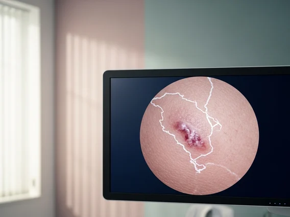
Clark Level IV skin cancer refers to a specific classification used in the histological assessment of melanoma, indicating the depth of tumor invasion within the skin layers. Understanding this level is crucial for determining prognosis and guiding treatment strategies for patients.
Key Takeaways
- Clark Level IV melanoma definition indicates tumor invasion into the reticular dermis, signifying a more advanced stage of melanoma.
- This classification is based on histological depth, complementing the Breslow depth measurement for staging.
- The Clark Level 4 skin cancer prognosis is generally less favorable than earlier stages, necessitating aggressive treatment.
- Treatment typically involves surgical excision, often combined with adjuvant therapies like immunotherapy or targeted therapy.
- Regular follow-up and early detection remain vital for managing melanoma at any stage.
What is Clark Level IV Skin Cancer?
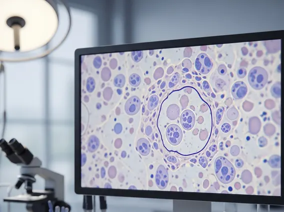
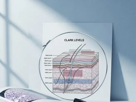
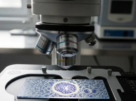
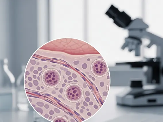
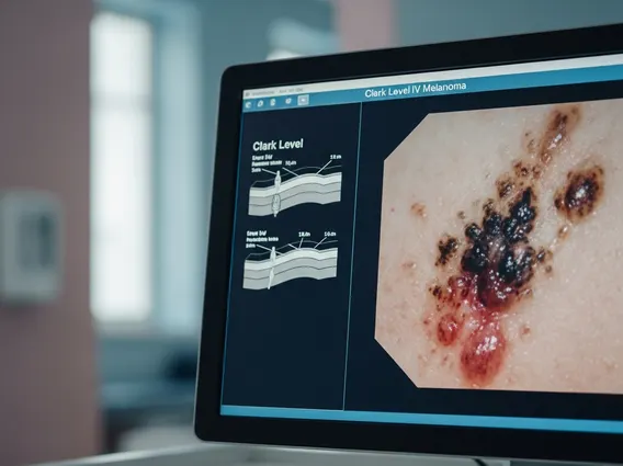
Clark Level IV melanoma definition refers to a histological classification system that describes the depth of invasion of melanoma cells into the layers of the skin. Developed by Dr. Wallace Clark, this system categorizes melanoma into five levels based on microscopic examination. Specifically, Clark Level IV indicates that the melanoma cells have invaded through the papillary dermis and into the reticular dermis. This layer is deeper than the papillary dermis and contains thicker collagen bundles and elastic fibers, signifying a more advanced stage of local invasion compared to Clark Levels I, II, and III.
While the Clark Level system provides valuable information about the anatomical depth of invasion, it is often used in conjunction with the Breslow depth measurement, which quantifies the tumor’s thickness in millimeters from the granular layer of the epidermis to the deepest point of invasion. Most modern staging systems, such as the American Joint Committee on Cancer (AJCC) staging, primarily rely on Breslow depth due to its stronger prognostic value, but Clark Level IV can still offer additional context regarding the tumor’s biological behavior and potential for metastasis.
Prognosis for Clark Level IV Melanoma
The Clark Level 4 skin cancer prognosis is generally considered less favorable compared to melanomas classified at earlier Clark Levels. This is because deeper invasion into the reticular dermis suggests a higher likelihood of the cancer cells having reached blood vessels or lymphatic channels, increasing the risk of regional lymph node involvement and distant metastasis. While Clark Level IV alone does not determine the entire prognosis, it is an important factor when combined with other elements like Breslow depth, ulceration, mitotic rate, and lymph node status.
For instance, a melanoma with a Clark Level IV invasion often correlates with a Breslow depth greater than 1.0 mm, which is a significant indicator of a poorer prognosis. According to data from various cancer registries, including those supported by organizations like the National Cancer Institute, the five-year survival rates for melanoma decrease as the stage advances. For localized melanoma (Stage I/II), survival rates are high, but they decline significantly for regional (Stage III) and distant (Stage IV) disease. While specific survival rates vary widely based on the overall AJCC stage, a Clark Level IV designation typically places a patient’s melanoma into a higher risk category, emphasizing the need for comprehensive treatment and diligent follow-up.
Clark Level IV Melanoma Treatment Options
Treatment for Clark Level IV melanoma is typically aggressive and multidisciplinary, aiming to remove the primary tumor and address any potential spread. The initial and most crucial step is wide local excision of the primary tumor, ensuring clear margins around the cancerous tissue. The extent of the surgical margin depends on the Breslow depth of the melanoma.
Beyond surgical removal of the primary lesion, additional treatments may include:
- Sentinel Lymph Node Biopsy (SLNB): Often recommended for melanomas with a Breslow depth greater than 0.8 mm or those with ulceration, to determine if cancer cells have spread to regional lymph nodes. If the sentinel node is positive, further lymph node dissection may be considered.
- Adjuvant Therapy: For patients with high-risk localized melanoma (e.g., thick tumors, ulceration, positive lymph nodes), adjuvant therapies are often used after surgery to reduce the risk of recurrence. These may include:
- Immunotherapy: Drugs like PD-1 inhibitors (e.g., pembrolizumab, nivolumab) or CTLA-4 inhibitors (e.g., ipilimumab) can stimulate the body’s immune system to fight cancer cells.
- Targeted Therapy: For melanomas with specific genetic mutations (e.g., BRAF mutation), targeted drugs (e.g., dabrafenib, vemurafenib, trametinib) can block the growth of cancer cells.
- Radiation Therapy: May be used in specific situations, such as to treat lymph nodes that cannot be surgically removed or to manage symptoms of metastatic disease.
- Clinical Trials: Patients may also be eligible for clinical trials exploring new treatments or combinations of therapies.
The choice of treatment plan is highly individualized, depending on the complete staging information, the patient’s overall health, and the presence of specific genetic markers. Close monitoring with regular follow-up appointments, including skin exams and imaging, is essential to detect any recurrence or new melanomas early.