Clark Level Iii Skin Cancer
Clark Level III Skin Cancer refers to a specific classification used in the histopathological assessment of melanoma, indicating the depth of tumor invasion into the skin layers. Understanding this classification is crucial for determining prognosis and guiding appropriate treatment strategies for patients.
Key Takeaways
- Clark Level III indicates melanoma cells have invaded the papillary dermis but have not reached the reticular dermis.
- This classification is based on histological depth, distinct from Breslow thickness, which measures vertical depth in millimeters.
- Early and accurate Clark Level III melanoma diagnosis is vital for effective management.
- Treatment typically involves surgical excision, with potential for further therapies depending on other factors.
- The Clark Level 3 skin cancer outlook is generally favorable compared to higher Clark levels, but regular follow-up is essential.
What is Clark Level III Skin Cancer?
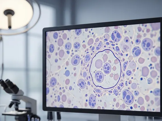
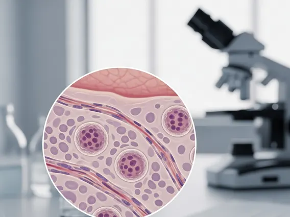
Clark Level III Skin Cancer describes a stage of melanoma where the malignant cells have invaded through the epidermis and into the papillary dermis, which is the superficial layer of the dermis. However, the tumor cells have not yet extended into the deeper reticular dermis. This classification system, developed by Dr. Wallace Clark, categorizes melanoma based on the anatomical depth of invasion, providing a histological snapshot of how far the cancer has spread within the skin layers.
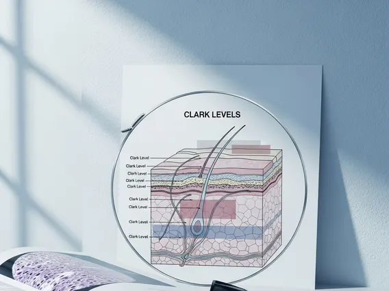
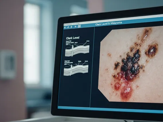
It is important to distinguish Clark levels from Breslow thickness. While both measure tumor depth, Breslow thickness provides a precise measurement in millimeters from the granular layer of the epidermis to the deepest point of tumor invasion, and it is generally considered a more significant prognostic factor than Clark level. Clark levels, on the other hand, offer a qualitative assessment of invasion relative to specific skin structures. For instance, Clark Level I is confined to the epidermis (in situ), Level II invades the papillary dermis but doesn’t fill it, Level III fills the papillary dermis, Level IV invades the reticular dermis, and Level V invades the subcutaneous fat.
Diagnosing and Treating Clark Level III Melanoma
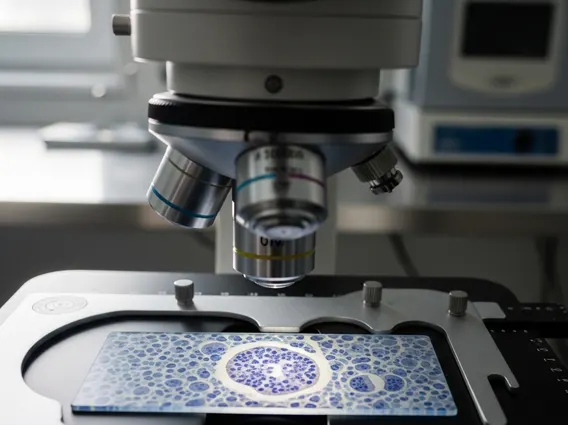
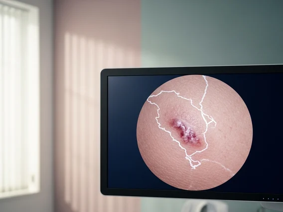
The Clark Level III melanoma diagnosis begins with a thorough clinical examination of suspicious skin lesions, followed by an excisional biopsy. During the biopsy, the entire lesion is removed and sent for histopathological analysis. A dermatopathologist examines the tissue under a microscope to determine the presence of melanoma, its specific characteristics, and its Clark level and Breslow thickness.
Once a diagnosis of Clark Level III melanoma is confirmed, the primary approach for Clark Level III melanoma treatment is surgical removal. This typically involves wide local excision, where the surgeon removes the original biopsy site along with a margin of healthy surrounding tissue to ensure all cancer cells are eradicated. The size of this margin depends on the Breslow thickness of the tumor. For melanomas with a Breslow thickness less than 1.0 mm, a 1 cm margin is often sufficient. In some cases, particularly if the Breslow thickness is greater than 1.0 mm, a sentinel lymph node biopsy may be considered to check for spread to nearby lymph nodes, although this is less common for lower-risk Clark Level III lesions unless other high-risk features are present.
Additional treatments, such as adjuvant therapy (e.g., immunotherapy or targeted therapy), are generally not required for localized Clark Level III melanoma unless there are signs of lymph node involvement or other high-risk features. Regular follow-up appointments are crucial to monitor for recurrence or the development of new lesions.
Clark Level III Melanoma Outlook and Prognosis
The Clark Level 3 skin cancer outlook is generally considered favorable, especially when compared to higher Clark levels (IV and V) or thicker Breslow measurements. The prognosis for melanoma is strongly correlated with the depth of invasion and whether the cancer has spread to lymph nodes or distant sites. Since Clark Level III indicates invasion into the papillary dermis but not beyond, it suggests a relatively early stage of the disease.
Survival rates for localized melanoma (which typically includes Clark Level III) are high. According to the American Cancer Society, the 5-year survival rate for localized melanoma is approximately 99% (American Cancer Society, 2023). However, it is important to remember that individual prognosis can vary based on several factors, including:
- Breslow thickness (the most significant prognostic factor)
- Presence or absence of ulceration
- Mitotic rate (how quickly cells are dividing)
- Patient’s age and overall health
- Location of the melanoma on the body
Patients with Clark Level III melanoma require diligent follow-up care, including regular skin exams, to detect any potential recurrence or new primary melanomas early. While alternative or complementary therapies may offer supportive care, they should never replace standard medical treatment for melanoma.