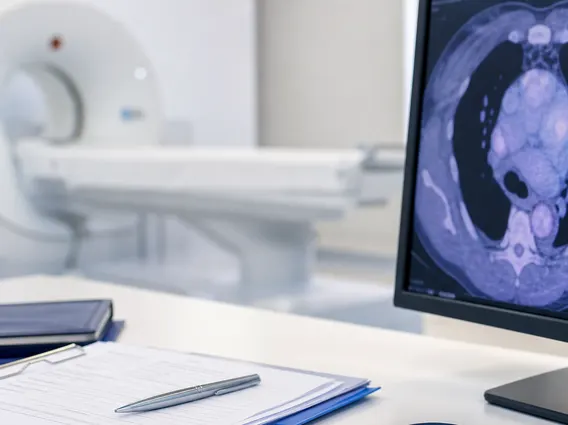
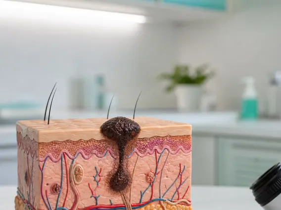
Chondrocyte
Chondrocytes are specialized cells crucial for the health and function of cartilage, a vital connective tissue in the body. Understanding these cells is fundamental to comprehending joint mechanics and various musculoskeletal conditions.
Key Takeaways
- Chondrocytes are the sole cells found within cartilage, responsible for its maintenance and repair.
- They produce and maintain the extracellular matrix (ECM) of cartilage, which provides its unique properties.
- These cells are essential for joint flexibility, shock absorption, and smooth movement.
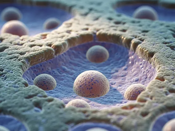
- Chondrocytes are primarily located within lacunae, small cavities embedded within the cartilage matrix.
- Their limited regenerative capacity makes cartilage injuries challenging to heal effectively.
What is a Chondrocyte? Definition and Primary Role
A Chondrocyte is the only cell type found within healthy cartilage, a specialized form of connective tissue that provides structural support and flexibility throughout the body. These cells are primarily responsible for producing and maintaining the cartilaginous matrix, which is composed predominantly of collagen fibers (especially type II), proteoglycans (like aggrecan), and elastin fibers. This unique extracellular matrix (ECM) gives cartilage its characteristic strength, elasticity, and remarkable ability to withstand compressive forces without deforming permanently. The primary role of chondrocytes is to ensure the structural integrity and functional capacity of cartilage, which is vital for smooth joint movement and shock absorption.
Chondrocytes originate from mesenchymal stem cells and undergo a complex process of differentiation to become mature cartilage cells. Their metabolic activity, though relatively low compared to other cell types, is crucial for the continuous turnover and repair of the cartilage matrix. However, their regenerative capacity is significantly limited, particularly in adult tissues. This inherent limitation in self-repair makes cartilage especially vulnerable to injury, wear-and-tear, and degenerative conditions such as osteoarthritis, where the breakdown of the matrix often outpaces the chondrocytes’ ability to repair it. Maintaining healthy chondrocytes is therefore paramount for long-term joint health and overall musculoskeletal function.
Chondrocyte Function and Where They Are Found
The fundamental chondrocyte function is centered on the synthesis and maintenance of the extracellular matrix (ECM) that gives cartilage its unique biomechanical properties. This involves the continuous production of key macromolecules, including various types of collagen (predominantly type II, which provides tensile strength) and large aggregating proteoglycans such as aggrecan (which traps water, providing resistance to compression). These components are critical for cartilage’s ability to act as a resilient shock absorber and to provide a smooth, low-friction surface that enables effortless movement within synovial joints. Without the constant and precise activity of chondrocytes, the cartilage matrix would degrade, leading to severe joint pain, stiffness, and ultimately, dysfunction.
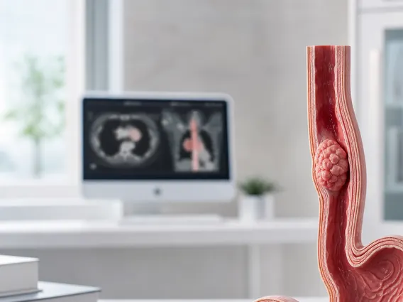
Where are chondrocytes found? These specialized cells are exclusively located within cartilage tissue, which is distributed in several crucial areas throughout the human body. They reside in small, fluid-filled cavities known as lacunae, which are embedded directly within the dense cartilage matrix. Cartilage itself is present in various forms and locations, each serving specific mechanical roles:
- Articular cartilage: Covers the ends of bones within synovial joints (e.g., knees, hips, shoulders), facilitating smooth articulation and distributing loads across the joint surfaces.
- Tracheal and bronchial rings: Provide essential structural support to keep the airways open, preventing collapse during breathing.
- External ear and nasal septum: Contribute to the flexible shape and structural integrity of these facial features.
- Intervertebral discs: Form resilient cushions between the vertebrae in the spine, absorbing shock and allowing spinal flexibility.
- Costal cartilage: Connects the ribs to the sternum, allowing for chest wall expansion during respiration.
Cartilage is notably avascular, meaning it lacks a direct blood supply, and also aneural (lacking nerves) and alymphatic (lacking lymphatic vessels). Consequently, chondrocytes receive their nutrients and dispose of metabolic waste products primarily through diffusion from the surrounding synovial fluid (in joints) or from the perichondrium, a dense connective tissue layer that surrounds most cartilage. This avascular nature contributes to the relatively slow metabolic rate of chondrocytes and significantly limits their capacity for self-repair after injury, underscoring the importance of their continuous, albeit slow, matrix maintenance activities.