Barium Meal Photofluorography
Barium Meal Photofluorography is a specialized medical imaging technique used to visualize and assess the upper gastrointestinal (GI) tract. This diagnostic procedure plays a crucial role in identifying various conditions affecting the esophagus, stomach, and duodenum.
Key Takeaways
- Barium Meal Photofluorography is an X-ray imaging technique for the upper GI tract.
- It uses a barium contrast agent to highlight internal structures.
- The procedure helps diagnose conditions like ulcers, strictures, and tumors.
- Patients typically fast before the test and drink a barium solution during imaging.
What is Barium Meal Photofluorography?
Barium Meal Photofluorography refers to a diagnostic imaging method that captures a series of X-ray images of the upper digestive system. This technique, also known as a barium swallow or upper GI series, involves the patient drinking a liquid containing barium sulfate, a radiopaque contrast agent. As the barium travels through the esophagus, stomach, and duodenum, it coats the lining of these organs, making them visible on X-ray images. The process allows medical professionals to observe the shape, function, and integrity of these structures, providing valuable insights into potential abnormalities. Essentially, barium meal photofluorography explained is a dynamic X-ray examination that helps in the detection of various gastrointestinal disorders.
Barium Meal Photofluorography Procedure and How It Works
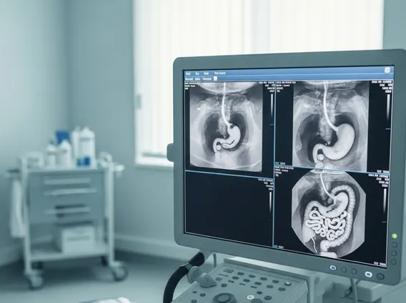
The Barium Meal Photofluorography procedure begins with patient preparation, which typically involves fasting for several hours prior to the examination to ensure the upper GI tract is empty. This allows for clearer visualization of the mucosal lining. During the test, the patient is asked to drink a thick, chalky liquid containing barium sulfate. The radiologist then uses a specialized X-ray machine to take a rapid sequence of images as the barium moves through the esophagus, stomach, and into the small intestine. The patient may be asked to change positions or swallow at specific times to help coat the organs thoroughly and capture different views.
The mechanism of how it works relies on the properties of barium sulfate. Barium is opaque to X-rays, meaning it absorbs X-ray radiation more effectively than surrounding soft tissues. When ingested, it coats the inner walls of the upper GI tract, creating a clear outline of the organs. This allows the radiologist to identify any irregularities in the mucosal lining, such as ulcers, polyps, strictures, or tumors, which would otherwise be invisible on standard X-rays. The rapid imaging captures the dynamic process of swallowing and digestion, providing functional information in addition to structural details.
Indications for Barium Meal Photofluorography
Barium meal photofluorography indications include a range of symptoms and suspected conditions affecting the upper gastrointestinal tract. This diagnostic tool is often employed when patients present with symptoms that suggest an issue in the esophagus, stomach, or duodenum. It helps clinicians pinpoint the cause of discomfort or dysfunction, guiding appropriate treatment strategies.
Common reasons for performing this test include:
- Persistent Dysphagia: Difficulty or pain when swallowing, which may indicate strictures, tumors, or motility disorders in the esophagus.
- Chronic Heartburn or Acid Reflux: To assess for gastroesophageal reflux disease (GERD), hiatal hernias, or complications like Barrett’s esophagus.
- Unexplained Abdominal Pain: Especially pain localized to the upper abdomen, which could be indicative of ulcers, inflammation, or other gastric pathologies.
- Nausea and Vomiting: When the cause is unclear and other diagnostic methods have not provided a definitive answer.
- Weight Loss: Unintentional weight loss that may be associated with malabsorption, tumors, or other GI conditions affecting nutrient intake or digestion.
- Suspected Ulcers or Tumors: To visualize and confirm the presence of gastric or duodenal ulcers, polyps, or cancerous growths.
By providing detailed images of the upper GI tract, Barium Meal Photofluorography assists in the accurate diagnosis and management of these and other related conditions.