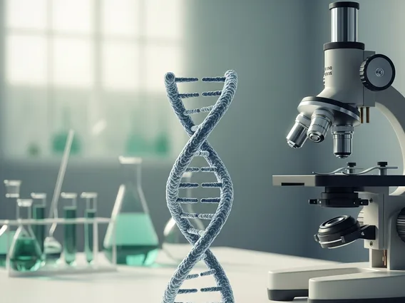
Histology
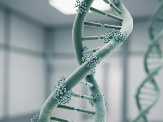
Histology is a fundamental branch of biology and medicine dedicated to the microscopic study of biological tissues. It provides crucial insights into the structure and organization of cells and tissues, essential for understanding both normal physiological processes and disease pathology.
Key Takeaways
- Histology is the microscopic study of tissues, vital for understanding biological structures and functions.
- It involves preparing and examining tissue samples to diagnose diseases and research biological processes.
- The human body is composed of four primary tissue types: epithelial, connective, muscle, and nervous tissue.
- Understanding tissue organization and cellular morphology is key to a comprehensive Histology study guide.
- Histology basics explained include tissue processing, staining techniques, and microscopic analysis.
What is Histology?
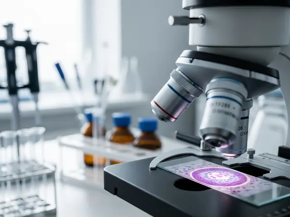
Histology refers to the scientific discipline focused on the detailed microscopic examination of biological tissues. This field is indispensable in medicine, serving as a cornerstone for diagnosing diseases, understanding their progression, and guiding treatment strategies. Through histology, scientists and clinicians can observe the intricate cellular architecture, tissue organization, and extracellular components that constitute organs and systems within the body.
The process typically involves taking tissue samples (biopsies), which are then meticulously prepared through fixation, processing, embedding, sectioning into very thin slices, and finally staining. Staining techniques, such as Hematoxylin and Eosin (H&E), are critical as they highlight different cellular and tissue components, making them visible under a light microscope. This detailed visualization allows for the identification of abnormalities that may indicate various conditions, from infections and inflammatory responses to cancerous growths.
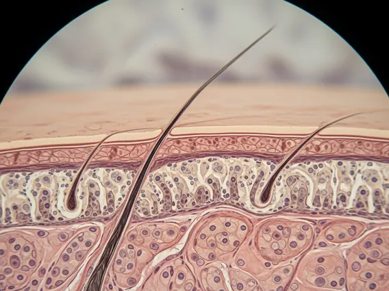
Types of Tissues in Histology
The human body is organized into four primary types of tissues, each with distinct structures and functions that are thoroughly explored in Histology. Understanding these fundamental categories is essential for any comprehensive Types of tissues histology study, as they form the basis of all organs and systems.
- Epithelial Tissue: This tissue forms coverings for body surfaces, lines body cavities and hollow organs, and constitutes glands. Its functions include protection, secretion, absorption, and filtration. Examples include the epidermis of the skin and the lining of the digestive tract.
- Connective Tissue: Characterized by its diverse forms and functions, connective tissue supports, connects, and protects other tissues and organs. It includes bone, cartilage, fat, blood, and fibrous connective tissues. Its primary role is to bind structures together, provide support, and transport substances.
- Muscle Tissue: Specialized for contraction, muscle tissue is responsible for movement. There are three types: skeletal muscle (voluntary movement), cardiac muscle (pumping blood in the heart), and smooth muscle (involuntary movements in internal organs).
- Nervous Tissue: Comprising the brain, spinal cord, and nerves, nervous tissue is responsible for transmitting electrical signals throughout the body. It allows for communication between different body parts, coordinating actions and responses to stimuli.
Each tissue type exhibits unique cellular arrangements and extracellular matrix compositions, which are meticulously analyzed during histological examination to discern normal physiology from pathological states.
Histology Basics: A Study Guide
For anyone embarking on a Histology study guide, grasping the foundational principles and methodologies is paramount. This field requires a keen eye for detail and an understanding of how tissue preparation affects microscopic appearance. A solid grasp of Histology basics explained includes familiarity with the steps involved in preparing tissue samples for examination, as well as the ability to identify different cell types and tissue structures under a microscope.
Key areas of focus for a histology student typically include:
- Tissue Processing: Understanding the steps from fixation (preserving tissue structure) to dehydration, clearing, and embedding in paraffin wax.
- Sectioning: Learning how microtomes are used to cut extremely thin slices of embedded tissue.
- Staining Techniques: Familiarity with common stains like H&E, and specialized stains that highlight specific components (e.g., Masson’s trichrome for collagen, PAS for carbohydrates).
- Microscopic Anatomy: Developing the skill to identify the four basic tissue types and their subtypes, as well as the cellular components within them, using a light microscope.
- Pathological Correlates: Recognizing how normal tissue architecture deviates in various disease states, which is a critical application of histological knowledge in clinical diagnostics.
Mastering these basics provides a strong foundation for advanced studies in pathology, medical research, and clinical practice, enabling accurate interpretation of tissue samples for diagnostic and investigative purposes.