Histologic Examination
Histologic Examination is a fundamental diagnostic procedure in medicine, involving the microscopic study of tissues to detect and characterize diseases. It plays a critical role in confirming diagnoses, guiding treatment, and understanding disease progression.
Key Takeaways
- Histologic Examination involves the detailed microscopic analysis of tissue samples.
- Its primary purpose is to accurately diagnose diseases, particularly cancers, and inform treatment strategies.
- The process includes tissue collection, preparation (fixation, embedding, sectioning, staining), and microscopic evaluation by a pathologist.
- This examination is crucial for confirming diagnoses, assessing disease severity, and predicting patient outcomes.
What is Histologic Examination?
Histologic Examination refers to the microscopic study of tissues to observe their structure, composition, and any abnormalities. This essential diagnostic tool involves preparing tissue samples obtained from biopsies or surgical resections, which are then thinly sliced, stained, and examined under a microscope by a pathologist. The detailed analysis allows for the identification of cellular changes, tissue architecture disruptions, and the presence of foreign substances, all of which are critical for diagnosing a wide range of medical conditions. This comprehensive approach ensures that the underlying pathology of a disease is thoroughly understood, providing crucial insights into its nature and progression. The process, often referred to as histology examination explained, provides definitive answers regarding the nature of a disease, from inflammatory conditions and infections to benign growths and malignant tumors, forming the cornerstone of many diagnostic pathways in modern medicine.
Purpose of Histologic Examination
The primary purpose of Histologic Examination is to provide a definitive diagnosis of various diseases, especially those involving abnormal tissue growth or cellular changes. Pathologists meticulously analyze tissue samples to:
- Confirm or rule out a suspected diagnosis, offering clarity where clinical findings alone may be inconclusive.
- Determine the precise type and stage of cancer, which is absolutely vital for developing an effective treatment plan. For instance, according to the World Health Organization (WHO), cancer is a leading cause of death globally, and accurate histologic diagnosis is paramount for effective management and improving patient survival rates.
- Assess the severity and extent of inflammatory or infectious diseases, guiding appropriate therapeutic interventions.
- Evaluate the effectiveness of medical treatments by comparing tissue samples taken before and after therapy, allowing for adjustments if necessary.
- Identify the presence of specific molecular markers or characteristics within the tissue that can predict disease behavior or a patient’s response to certain targeted drugs.
- Aid in forensic investigations by analyzing tissue damage or the presence of foreign materials, contributing to legal and medical understanding.
This detailed tissue analysis is indispensable for guiding clinical decisions, personalizing patient care, and ultimately improving health outcomes across numerous medical specialties.
The Histologic Examination Process
The process of Histologic Examination is a meticulous multi-step procedure designed to preserve tissue integrity and enable detailed microscopic analysis. It typically begins with the collection of a tissue sample, followed by a precise sequence of laboratory steps to prepare the specimen for examination:
- Tissue Collection: A clinician performs a biopsy or surgical excision to obtain the tissue specimen.
- Fixation: The collected tissue is immediately placed in a fixative, commonly formalin, to halt decomposition and preserve cellular structures.
- Gross Examination: A pathologist or assistant macroscopically examines the fixed tissue, describing its appearance and selecting areas for microscopic analysis.
- Tissue Processing: The selected tissue undergoes dehydration (alcohol), clearing (xylene), and infiltration with molten paraffin wax.
- Embedding: The processed tissue is oriented and embedded into a block of paraffin wax, providing support for sectioning.
- Sectioning: Using a microtome, extremely thin slices (typically 3-5 micrometers) of the tissue block are cut and mounted on glass slides.
- Staining: Slides are stained with various dyes, such as hematoxylin and eosin (H&E), to highlight cellular components and tissue structures.
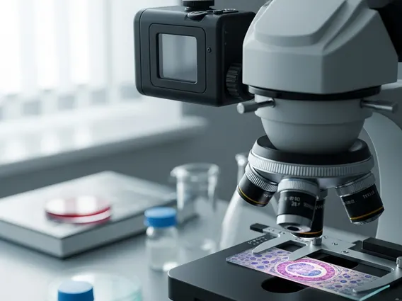
- Microscopic Examination: A qualified pathologist examines the stained slides under a microscope, identifying abnormalities, diagnosing diseases, and preparing a comprehensive report for the referring clinician.
This systematic approach ensures that high-quality slides are produced, enabling accurate and reliable diagnostic interpretations crucial for patient care.