Mri
Magnetic Resonance Imaging (Mri) is a sophisticated medical imaging technique that provides detailed pictures of organs, soft tissues, bone, and virtually all other internal body structures. It is a vital diagnostic tool in modern medicine, aiding in the detection and monitoring of a wide range of conditions.
Key Takeaways
- Mri utilizes powerful magnets and radio waves to generate highly detailed images of the body’s internal structures.
- Unlike X-rays or CT scans, Mri does not use ionizing radiation, making it a safe option for repeated imaging.
- It is particularly effective for visualizing soft tissues, including the brain, spinal cord, muscles, ligaments, and internal organs.
- The procedure is non-invasive, requiring patients to lie still inside a scanner while images are captured.
- Mri plays a crucial role in diagnosing conditions ranging from neurological disorders and musculoskeletal injuries to cancer and cardiovascular diseases.
What is an MRI (Magnetic Resonance Imaging) Scan?
An Mri scan, or Magnetic Resonance Imaging scan, is a non-invasive medical imaging test that uses a strong magnetic field and radio waves to create detailed images of organs and tissues within the body. It is distinct from X-rays and CT scans because it does not employ ionizing radiation, making it a preferred choice for certain diagnostic situations, especially for repeated imaging or in sensitive areas.
Magnetic Resonance Imaging (MRI) refers to the technology that harnesses the natural magnetic properties of the body’s hydrogen atoms. These atoms, abundant in water and fat, respond to the magnetic field and radio waves, allowing a computer to construct cross-sectional images. This capability makes Mri invaluable for examining soft tissues that are often difficult to visualize with other imaging methods.
How MRI Technology Works
The fundamental principle behind how Mri technology works involves the manipulation of hydrogen atoms, which are plentiful in the water molecules found throughout the body. When a patient enters an Mri scanner, a powerful magnetic field causes these hydrogen protons to align in the same direction. Short bursts of radio waves are then emitted, temporarily knocking these aligned protons out of alignment.
When the radio waves are turned off, the hydrogen protons quickly realign with the main magnetic field, releasing energy in the form of radio signals. Different tissues release these signals at varying rates and intensities, which are detected by the Mri scanner’s receiver. A sophisticated computer then processes these signals, translating them into detailed, high-resolution images of the body’s internal structures. This intricate process allows clinicians to differentiate between healthy and diseased tissue with remarkable clarity.
Uses and Procedure of an MRI Scan
The uses of Mri in medicine are extensive, making it a versatile diagnostic tool across many specialties. It is particularly adept at visualizing soft tissues, which include the brain, spinal cord, nerves, muscles, ligaments, and tendons. Mri scans are routinely used to diagnose and monitor conditions such as:
- Neurological disorders (e.g., strokes, tumors, multiple sclerosis, brain injuries)
- Musculoskeletal injuries (e.g., torn ligaments, cartilage damage, bone infections)
- Cancers (for detection, staging, and monitoring treatment response in various organs)
- Cardiovascular issues (e.g., heart muscle damage, blood vessel abnormalities)
- Abdominal and pelvic conditions (e.g., liver disease, kidney issues, uterine fibroids)
According to the Organization for Economic Co-operation and Development (OECD), over 100 million Mri exams are performed annually across its member countries, highlighting its widespread adoption in healthcare for these diverse applications.
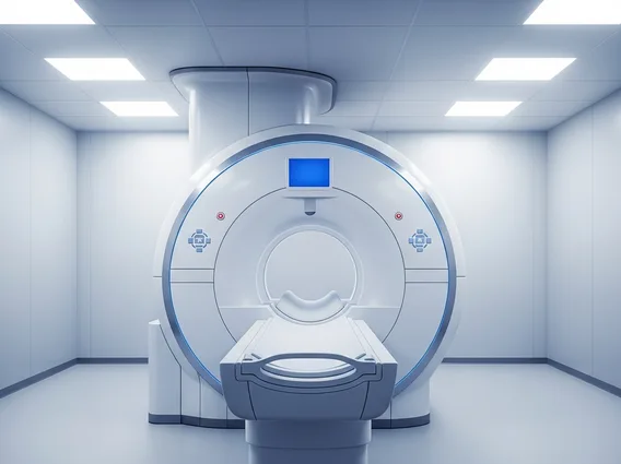
The Mri scan procedure explained typically involves the patient lying on a movable examination table that slides into a large, tube-shaped scanner. During the scan, it is crucial for the patient to remain very still to ensure clear images. The process can be noisy, so earplugs or headphones are often provided. Depending on the area being examined and the specific diagnostic needs, a contrast agent may be injected intravenously to enhance the visibility of certain tissues or blood vessels.
Preparation for an Mri scan usually involves removing all metal objects, such as jewelry, watches, and hearing aids, due to the powerful magnetic field. Patients with certain implanted medical devices, like pacemakers or some cochlear implants, may not be able to undergo an Mri. The duration of an Mri scan can vary from 15 minutes to over an hour, depending on the complexity of the examination.