Mammosite
Mammosite is a targeted radiation therapy used in the treatment of early-stage breast cancer. This approach delivers radiation directly to the tumor bed, aiming to reduce treatment time and minimize exposure to healthy tissues.
Key Takeaways
- Mammosite is a form of accelerated partial breast irradiation (APBI) for early-stage breast cancer.
- It utilizes a balloon catheter to deliver radiation internally to the lumpectomy cavity.
- The therapy aims to precisely target the tumor bed, sparing surrounding healthy breast tissue and vital organs.
- Treatment typically involves twice-daily sessions over five consecutive days.
- Common side effects are generally localized and may include skin irritation, bruising, or discomfort.
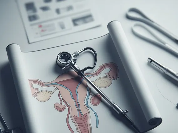
What is Mammosite?
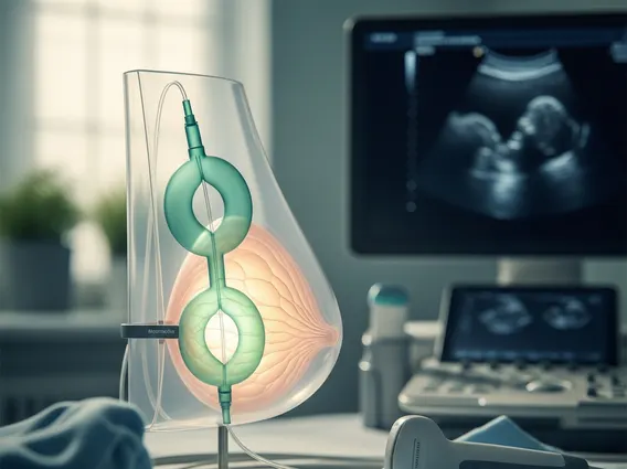
Mammosite refers to a specific type of balloon catheter-based brachytherapy system designed for accelerated partial breast irradiation (APBI) following a lumpectomy for early-stage breast cancer. This innovative approach allows for the precise delivery of radiation directly to the area where the tumor was removed, known as the tumor bed. By focusing radiation on this specific site, Mammosite therapy aims to destroy any remaining microscopic cancer cells while significantly reducing the radiation dose to the rest of the breast, chest wall, and nearby organs.
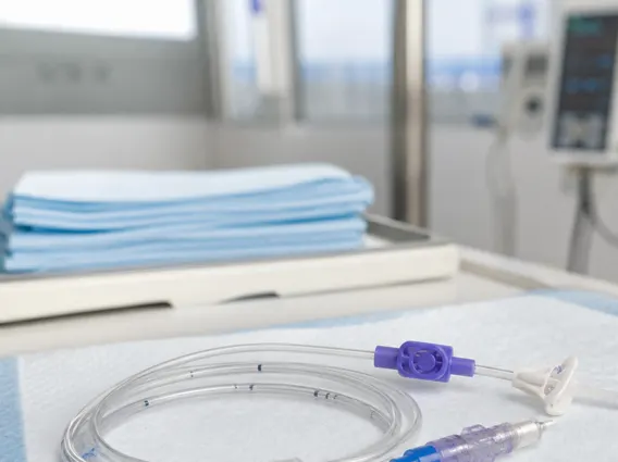
The system consists of a small, soft balloon catheter that is temporarily placed into the lumpectomy cavity. Once in position, the balloon is inflated with saline solution to conform to the cavity’s shape. This method represents a significant advancement in radiation oncology, offering a more convenient and often shorter treatment course compared to traditional whole breast radiation therapy for carefully selected patients.
How Mammosite Radiation Therapy Works for Breast Cancer
Mammosite radiation therapy is a localized treatment option for certain patients with early-stage breast cancer, often chosen as an alternative to traditional whole breast radiation. The process begins after a lumpectomy, where the cancerous tissue is surgically removed, leaving a cavity. A small balloon catheter is then inserted into this cavity. Once the catheter is properly positioned and the balloon is inflated, it creates a precise target for radiation delivery.
During the treatment sessions, a radioactive source is temporarily placed inside the balloon, delivering a high dose of radiation directly to the tissue immediately surrounding the lumpectomy cavity. This targeted delivery is a key aspect of mammosite breast cancer treatment, aiming to destroy any remaining microscopic cancer cells while significantly reducing radiation exposure to the rest of the breast, chest wall, and vital organs like the heart and lungs. The treatment typically involves twice-daily sessions over five consecutive days, making it a much shorter course than conventional radiation therapy, which can last several weeks. Clinical studies have indicated that APBI methods, including Mammosite, offer comparable local control rates to whole breast radiation therapy for appropriately selected patients with early-stage breast cancer, often with improved patient convenience and quality of life.
The procedure generally follows these steps:
- Catheter Insertion: After lumpectomy, a balloon catheter is surgically placed into the tumor bed.
- Balloon Inflation: The balloon is inflated with saline to fit the cavity, ensuring optimal contact with the surrounding tissue.
- Treatment Planning: Imaging (like CT scans) is used to verify catheter placement and plan the precise radiation dose.
- Radiation Delivery: A radioactive source is temporarily inserted into the balloon for short periods during each treatment session.
- Catheter Removal: After the prescribed course of radiation (typically five days), the balloon is deflated, and the catheter is removed.
Mammosite Radiation Therapy Side Effects
While generally well-tolerated, Mammosite side effects can occur, similar to other forms of radiation therapy, though often localized and less severe due to the targeted nature of the treatment. The most common side effects are typically confined to the treated breast and the area around the catheter insertion site.
Patients may experience temporary changes in the treated breast, such as redness, swelling, bruising, or discomfort at the catheter insertion site. Some individuals might also notice a feeling of tightness or tenderness in the breast. Fatigue is another possible side effect, though it is often less pronounced compared to whole breast radiation therapy. These effects are usually manageable with supportive care and tend to resolve within a few weeks or months after the completion of treatment.
Less common but potential side effects include infection at the catheter site, fluid accumulation (seroma) around the catheter, or fat necrosis (a benign hardening of fatty tissue) in the treated area. Serious complications are rare. Patients are closely monitored throughout the treatment period and follow-up to address any concerns and manage side effects effectively, ensuring the best possible outcome.