Magnetic Resonance Imaging
Magnetic Resonance Imaging (MRI) is a powerful non-invasive diagnostic tool used in medicine to produce detailed images of organs, soft tissues, bone, and virtually all other internal body structures. It plays a crucial role in detecting and diagnosing a wide range of conditions without using ionizing radiation.
Key Takeaways
- MRI uses strong magnetic fields and radio waves to create detailed internal body images.
- It is a non-invasive procedure that does not involve ionizing radiation, making it safe for repeated use.
- MRI scans are invaluable for diagnosing conditions affecting the brain, spinal cord, joints, and soft tissues.
- The procedure requires patients to remain still inside a scanner, which can be challenging for some.
- While generally safe, contraindications include certain metallic implants and claustrophobia.
What is Magnetic Resonance Imaging (MRI)?
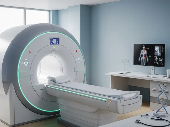
Magnetic Resonance Imaging (MRI) is an advanced medical imaging technique that utilizes a powerful magnetic field and radio waves to generate cross-sectional images of organs and soft tissues within the body. Unlike X-rays or CT scans, MRI does not use ionizing radiation, making it a safer option for repeated examinations, especially for vulnerable populations. The detailed images produced by MRI allow healthcare professionals to accurately diagnose a variety of medical conditions affecting the brain, spinal cord, heart, bones, joints, and other internal structures.
How Does Magnetic Resonance Imaging Work?
An MRI scan operates on the principle of nuclear magnetic resonance. When a patient enters the MRI scanner, their body is placed within a strong magnetic field. This field causes the protons within the water molecules in the body’s tissues to align with the magnetic field. A radiofrequency current is then briefly pulsed through the patient, knocking these aligned protons out of alignment. When the radiofrequency pulse is turned off, the protons relax back into alignment with the main magnetic field, releasing energy in the form of radio signals. Different tissues relax at different rates and emit signals of varying intensity. These signals are detected by the MRI scanner’s antenna and sent to a computer, which processes them to create detailed, cross-sectional images of the body. The computer can then reconstruct these signals into two-dimensional slices or even three-dimensional models, allowing for comprehensive visualization of internal structures.
Uses, Benefits, and Risks of MRI Scans
The **uses of MRI in medical diagnosis** are extensive, covering nearly every part of the body. It is particularly effective for imaging soft tissues, making it indispensable for neurological conditions, musculoskeletal injuries, and detecting tumors. Common applications include:
- Brain and Spinal Cord: Diagnosing strokes, tumors, aneurysms, multiple sclerosis, and spinal cord injuries.
- Joints and Bones: Evaluating torn ligaments, cartilage damage, bone infections, and arthritis.
- Internal Organs: Assessing conditions in the heart, liver, kidneys, pancreas, and reproductive organs.
- Breast Imaging: Used as a supplementary tool for breast cancer screening, especially for high-risk individuals.
The **benefits and risks of MRI** scans are important considerations for patients and clinicians. Benefits include its non-invasive nature, the absence of ionizing radiation, superior soft tissue contrast, and versatility in imaging various body parts. Potential risks and limitations include contraindications for patients with certain metallic implants due to the strong magnetic field, the possibility of claustrophobia in the enclosed scanner, and the loud noises produced during the scan. While generally safe, some patients may experience rare allergic reactions to gadolinium-based contrast agents. The decision to undergo an MRI scan is made by a healthcare provider, weighing these factors against the diagnostic information it can provide.