Magnetic Resonance Cholangiopancreatography
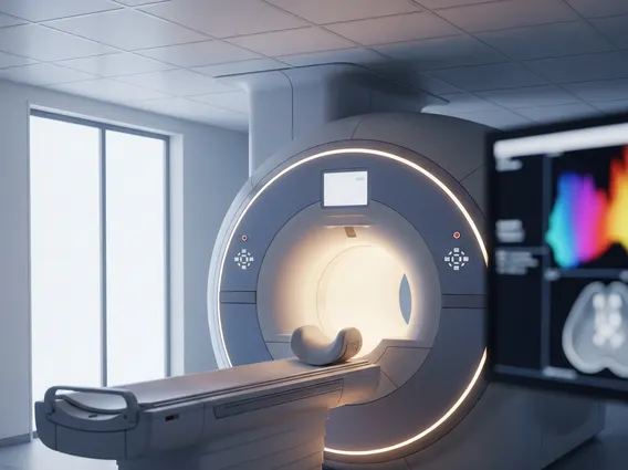
Magnetic Resonance Cholangiopancreatography (MRCP) is a non-invasive medical imaging technique that utilizes magnetic resonance imaging (MRI) to visualize the bile ducts, pancreatic duct, and gallbladder. This advanced diagnostic tool provides detailed images without the need for intravenous contrast agents or ionizing radiation.
Key Takeaways
- MRCP is a specialized MRI scan that creates detailed images of the bile and pancreatic ducts.
- It is a non-invasive procedure, meaning it does not require endoscopy or exposure to ionizing radiation.
- The technique relies on the high fluid content of the ducts to generate bright signals, making blockages or abnormalities clearly visible.
- MRCP is highly effective in diagnosing conditions such as gallstones, strictures, tumors, and inflammation of the bile and pancreatic systems.
- Its primary benefits include its non-invasive nature, lack of radiation, and ability to provide comprehensive anatomical information.
What is Magnetic Resonance Cholangiopancreatography (MRCP)?
Magnetic Resonance Cholangiopancreatography (MRCP) is a sophisticated imaging method that specifically targets the biliary and pancreatic ductal systems. This non-invasive diagnostic procedure uses a powerful magnetic field and radio waves to generate detailed cross-sectional images of the liver, gallbladder, bile ducts, pancreas, and pancreatic duct. The primary goal of an MRCP scan is to detect and characterize abnormalities within these fluid-filled structures, offering a comprehensive Magnetic Resonance Cholangiopancreatography explanation of their condition.
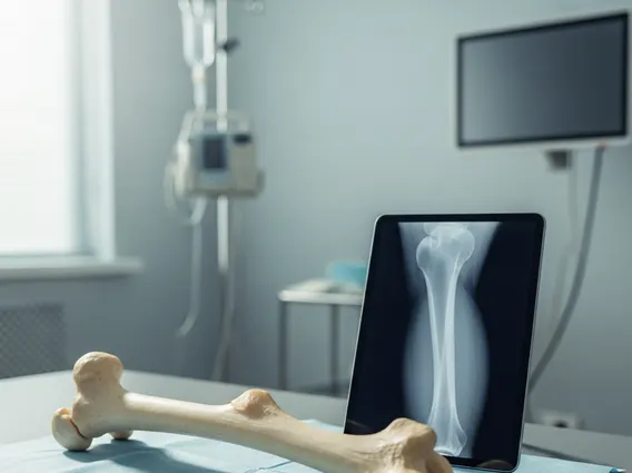
Unlike traditional X-rays or CT scans, MRCP does not involve ionizing radiation, making it a safer option for repeated examinations or for patients sensitive to radiation exposure. It is particularly useful for evaluating patients with symptoms such as jaundice, abdominal pain, or unexplained weight loss, which may indicate issues within the hepatobiliary or pancreatic systems. The procedure is generally well-tolerated and requires minimal preparation, typically involving fasting for a few hours prior to the scan.
Mechanism of MRCP Imaging
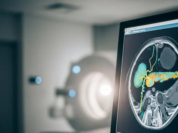
The fundamental principle behind how MRCP works lies in its ability to highlight static or slow-moving fluids within the body. During an MRCP scan, the MRI scanner uses strong magnetic fields and radiofrequency pulses to align the protons within the water molecules of the body’s tissues. When the radiofrequency pulse is turned off, these protons release energy, which is detected by the scanner and converted into detailed images.
For MRCP, a specific imaging sequence known as heavily T2-weighted imaging is employed. This sequence causes fluid-filled structures, such as the bile and pancreatic ducts, to appear very bright (hyperintense) on the images, while suppressing signals from surrounding solid organs and tissues. This contrast allows radiologists to clearly visualize the anatomy of the ducts and identify any blockages, dilations, strictures, or other abnormalities. The patient lies still on a table that slides into the MRI machine, and the scan typically takes between 20 to 45 minutes, during which they may be asked to hold their breath for short periods to minimize motion artifacts.
Clinical Applications and Benefits
MRCP has become an invaluable tool in gastroenterology and hepatology for diagnosing a wide array of conditions affecting the bile and pancreatic ducts. The MRCP scan uses and benefits are extensive, primarily due to its non-invasive nature and high diagnostic accuracy. It is frequently utilized to investigate the causes of obstructive jaundice, acute and chronic pancreatitis, and suspected tumors of the pancreas or bile ducts.
Key clinical applications include:
- Gallstones: Detecting stones within the bile ducts (choledocholithiasis) or gallbladder.
- Bile Duct Obstruction: Identifying strictures (narrowing), tumors, or other causes of blockage in the bile ducts.
- Pancreatic Duct Abnormalities: Visualizing stones, strictures, or cysts within the pancreatic duct, often associated with pancreatitis.
- Congenital Anomalies: Diagnosing birth defects of the bile or pancreatic ducts, such as choledochal cysts.
- Pre-surgical Planning: Providing detailed anatomical maps before surgical interventions on the biliary or pancreatic systems.
One of the significant benefits of MRCP is its ability to provide detailed images without the risks associated with more invasive procedures like Endoscopic Retrograde Cholangiopancreatography (ERCP). While ERCP is both diagnostic and therapeutic, it carries risks such as pancreatitis, perforation, and bleeding. MRCP offers a safe, non-invasive alternative for initial diagnosis, helping clinicians determine if a more invasive procedure is truly necessary. According to a study published in the journal Radiology, MRCP has a high sensitivity and specificity for detecting choledocholithiasis, often comparable to ERCP but with a significantly lower risk profile.