Nuclear Magnetic Resonance Imaging
Nuclear Magnetic Resonance Imaging (NMRI) is a powerful, non-invasive diagnostic tool that provides highly detailed images of organs, soft tissues, bone, and virtually all other internal body structures. It plays a crucial role in modern medicine by aiding in the diagnosis and monitoring of a wide range of conditions.
Key Takeaways
- Nuclear Magnetic Resonance Imaging (NMRI) is a non-invasive medical imaging technique that uses strong magnetic fields and radio waves to create detailed images of the body’s internal structures.
- It works by aligning the protons within the body’s water molecules using a powerful magnet, then briefly disrupting this alignment with radiofrequency pulses.
- As the protons realign, they release energy signals that are detected by the scanner and processed into cross-sectional images.
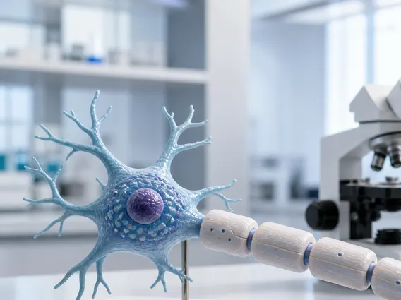
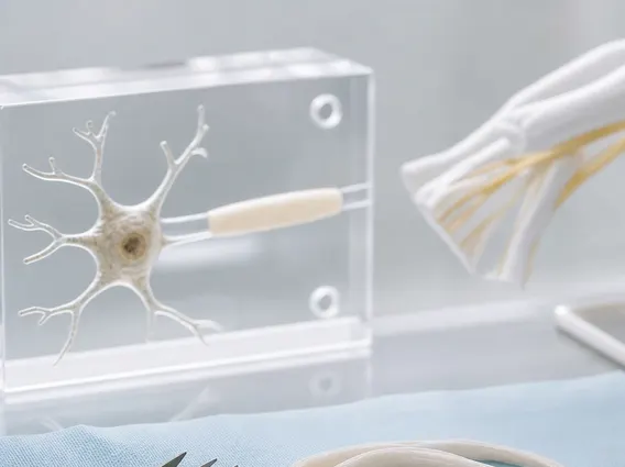
- NMRI is highly effective for visualizing soft tissues, making it invaluable for diagnosing conditions affecting the brain, spinal cord, joints, and internal organs.
- The term “nuclear” refers to the atomic nuclei (protons) involved, not to radioactivity, making it a safe procedure without ionizing radiation.
What is Nuclear Magnetic Resonance Imaging (NMRI)?
Nuclear Magnetic Resonance Imaging (NMRI) is an advanced medical imaging technique that utilizes a strong magnetic field and radio waves to generate detailed cross-sectional images of organs and soft tissues within the body. Often referred to simply as Magnetic Resonance Imaging (MRI), the term “nuclear” in its full name refers to the atomic nuclei (specifically, the hydrogen protons in water molecules) that respond to the magnetic field, not to nuclear radiation. This distinction is important, as NMRI does not involve ionizing radiation, making it a safe diagnostic option for many patients, including pregnant women and children, when medically indicated.
The underlying phenomenon, Nuclear Magnetic Resonance (NMR), involves the absorption and emission of energy by atomic nuclei in a magnetic field. In the context of medical imaging, this allows clinicians to visualize anatomical structures with exceptional clarity, differentiating between various types of soft tissues more effectively than other imaging modalities like X-rays or CT scans. This capability is vital for diagnosing a broad spectrum of conditions, from neurological disorders to musculoskeletal injuries and cancer.
How Does Nuclear Magnetic Resonance Imaging Work?
The fundamental process of how does nmr imaging work relies on the interaction between the body’s hydrogen atoms and powerful magnetic fields. The human body is primarily composed of water, which contains hydrogen atoms, each with a single proton. These protons act like tiny magnets, normally spinning randomly. When a patient enters an NMRI scanner, a very strong magnetic field causes these protons to align with the field, much like compass needles pointing north.
Next, the scanner emits brief radiofrequency pulses. These pulses temporarily knock the aligned protons out of alignment. When the radiofrequency pulse is turned off, the protons relax and realign with the main magnetic field, releasing energy in the form of radio signals. The time it takes for the protons to realign, and the amount of energy they release, varies depending on the type of tissue they are in. For example, water-rich tissues like cerebrospinal fluid behave differently from fatty tissues or bone.
These emitted signals are detected by receiver coils in the NMRI scanner. A computer then processes these signals, converting them into detailed, high-resolution images. The variations in signal strength and decay times allow for the precise differentiation of various tissues, enabling the visualization of subtle abnormalities. The principles of MRI scanning are thus based on detecting these minute differences in proton behavior to construct a comprehensive map of the body’s internal structures.
Here are the core steps involved:
- Magnetic Field Application: A powerful magnet aligns the hydrogen protons in the body.
- Radiofrequency Pulses: Brief radio waves are sent, knocking protons out of alignment.
- Signal Emission: Protons relax and realign, emitting radio signals.
- Signal Detection: Receiver coils detect these varying signals.
- Image Reconstruction: A computer processes signals into detailed images.
Clinical Uses of Nuclear Magnetic Resonance Imaging
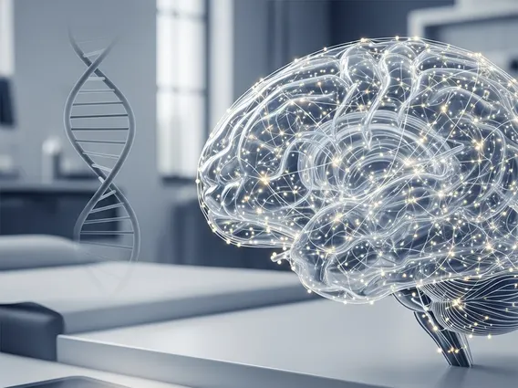
The uses of NMR in medicine are extensive and continue to expand due to its superior soft tissue contrast and non-ionizing nature. NMRI is particularly valuable for diagnosing conditions in areas where other imaging techniques may be less effective. For instance, it is the gold standard for imaging the brain and spinal cord, detecting conditions such as tumors, strokes, multiple sclerosis, and aneurysms. Its ability to visualize subtle changes in tissue composition makes it indispensable for neurological assessments.
Beyond neurological applications, NMRI is widely used for musculoskeletal imaging. It can clearly show ligaments, tendons, cartilage, and bone marrow, making it ideal for diagnosing sports injuries, arthritis, and disc problems in the spine. In oncology, NMRI helps in detecting, staging, and monitoring various cancers, including those of the breast, prostate, liver, and uterus, by providing detailed information about tumor size, location, and spread. According to the World Health Organization (WHO), medical imaging, including MRI, is critical for early diagnosis and treatment planning, significantly improving patient outcomes globally.
Furthermore, NMRI is employed in cardiovascular imaging to assess heart structure and function, detect blockages, and evaluate damage from heart attacks. It also plays a role in abdominal imaging for evaluating organs like the kidneys, liver, and pancreas. Its versatility and safety profile make it a cornerstone of modern diagnostic imaging, guiding treatment decisions and improving patient care across numerous medical specialties.